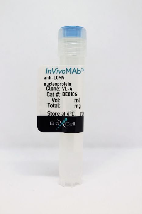
InVivoMAb anti-LCMV nucleoprotein
Product Details
The VL-4 antibody reacts with lymphocytic choriomeningitis virus (LCMV) nucleoprotein (NP), a 63 kDa structural protein. This antibody was generated by fusion of spleen cells of an LCMV strain WE immunized F1 rat with the YM3 myeloma cell line. This antibody has been shown to stain LCMV-infected cell internally with no surface staining. This antibody does not react with vaccinia, vesicular stomatitis or influenza virus-infected cells in the case of internal or surface staining.Specifications
| Isotype | Rat IgG2a, κ |
|---|---|
| Recommended Isotype Control(s) | InVivoMAb rat IgG2a isotype control, anti-trinitrophenol |
| Recommended Dilution Buffer | InVivoPure pH 7.0 Dilution Buffer |
| Conjugation | This product is unconjugated. Conjugation is available via our Antibody Conjugation Services. |
| Immunogen | LCMV strain WE |
| Reported Applications |
Immunofluorescence Flow cytometry |
| Formulation |
PBS, pH 7.0 Contains no stabilizers or preservatives |
| Endotoxin |
<2EU/mg (<0.002EU/μg) Determined by LAL gel clotting assay |
| Purity |
>95% Determined by SDS-PAGE |
| Sterility | 0.2 µm filtration |
| Production | Purified from cell culture supernatant in an animal-free facility |
| Purification | Protein G |
| RRID | AB_10949017 |
| Molecular Weight | 150 kDa |
| Storage | The antibody solution should be stored at the stock concentration at 4°C. Do not freeze. |
Recommended Products
-
Recommended Isotype Control(s)
InVivoMAb rat IgG2a isotype control, anti-trinitrophenol
-

Recommended Dilution Buffer
InVivoPure pH 7.0 Dilution Buffer
Flow Cytometry
Pritzl, C. J., et al. (2015). "A ceramide analogue stimulates dendritic cells to promote T cell responses upon virus infections" J Immunol 194(9): 4339-4349. PubMed
The ceramide family of lipids plays important roles in both cell structure and signaling in a diverse array of cell types, including immune cells. However, very little is known regarding how ceramide affects the activation of dendritic cells (DCs) in response to viral infection. In this study, we demonstrate that a synthetic ceramide analog (C8) stimulates DCs to increase the expansion of virus-specific T cells upon virus infection. Exogenously supplied C8 ceramide elevated the expression of DC maturation markers such as MHC class I and costimulatory molecules following infection with the clone 13 strain of lymphocytic choriomeningitis virus (LCMV) or influenza virus. Importantly, ceramide-conditioned, LCMV-infected DCs displayed an increased ability to promote expansion of virus-specific CD8(+) T cells when compared with virus-infected DCs. Furthermore, a locally instilled ceramide analog significantly increased virus-reactive T cell responses in vivo to both LCMV and influenza virus infections. Collectively, these findings provide new insights into ceramide-mediated regulation of DC responses against virus infection and help us establish a foundation for novel immune-stimulatory therapeutics.
Flow Cytometry
Ng, C. T., et al. (2015). "Blockade of interferon Beta, but not interferon alpha, signaling controls persistent viral infection" Cell Host Microbe 17(5): 653-661. PubMed
Although type I interferon (IFN-I) is thought to be beneficial against microbial infections, persistent viral infections are characterized by high interferon signatures suggesting that IFN-I signaling may promote disease pathogenesis. During persistent lymphocytic choriomeningitis virus (LCMV) infection, IFNalpha and IFNbeta are highly induced early after infection, and blocking IFN-I receptor (IFNAR) signaling promotes virus clearance. We assessed the specific roles of IFNbeta versus IFNalpha in controlling LCMV infection. While blockade of IFNbeta alone does not alter early viral dissemination, it is important in determining lymphoid structure, lymphocyte migration, and anti-viral T cell responses that lead to accelerated virus clearance, approximating what occurs during attenuation of IFNAR signaling. Comparatively, blockade of IFNalpha was not associated with improved viral control, but with early dissemination of virus. Thus, despite their use of the same receptor, IFNbeta and IFNalpha have unique and distinguishable biologic functions, with IFNbeta being mainly responsible for promoting viral persistence.
Flow Cytometry
Sullivan, B. M., et al. (2015). "Early virus-host interactions dictate the course of a persistent infection" PLoS Pathog 11(1): e1004588. PubMed
Many persistent viral infections are characterized by a hypofunctional T cell response and the upregulation of negative immune regulators. These events occur days after the initiation of infection. However, the very early host-virus interactions that determine the establishment of viral persistence remain poorly uncharacterized. Here we show that to establish persistence, LCMV must counteract an innate anti-viral immune response within eight hours after infection. While the virus triggers cytoplasmic RNA sensing pathways soon after infection, LCMV counteracts this pathway through a rapid increase in viral titers leading to a dysfunctional immune response characterized by a high cytokine and chemokine expression profile. This altered immune environment allows for viral replication in the splenic white pulp as well as infection of immune cells essential to an effective anti-viral immune response. Our findings illustrate how early events during infection critically dictate the characteristics of the immune response to infection and facilitate either virus control and clearance or persistence.
Immunofluorescence
Beura, L. K., et al. (2015). "Lymphocytic choriomeningitis virus persistence promotes effector-like memory differentiation and enhances mucosal T cell distribution" J Leukoc Biol 97(2): 217-225. PubMed
Vaccines are desired that maintain abundant memory T cells at nonlymphoid sites of microbial exposure, where they may be anatomically positioned for immediate pathogen interception. Here, we test the impact of antigen persistence on mouse CD8 and CD4 T cell distribution and differentiation by comparing responses to infections with different strains of LCMV that cause either acute or chronic infections. We used in vivo labeling techniques that discriminate between T cells present within tissues and abundant populations that fail to be removed from vascular compartments, despite perfusion. LCMV persistence caused up to approximately 30-fold more virus-specific CD8 T cells to distribute to the lung compared with acute infection. Persistent infection also maintained mucosal-homing alpha4beta7 integrin expression, higher granzyme B expression, alterations in the expression of the TRM markers CD69 and CD103, and greater accumulation of virus-specific CD8 T cells in the large intestine, liver, kidney, and female reproductive tract. Persistent infection also increased LCMV-specific CD4 T cell quantity in mucosal tissues and induced maintenance of CXCR4, an HIV coreceptor. This study clarifies the relationship between viral persistence and CD4 and CD8 T cell distribution and mucosal phenotype, indicating that chronic LCMV infection magnifies T cell migration to nonlymphoid tissues.
Flow Cytometry, Immunofluorescence
Herz, J., et al. (2015). "Therapeutic antiviral T cells noncytopathically clear persistently infected microglia after conversion into antigen-presenting cells" J Exp Med 212(8): 1153-1169. PubMed
Several viruses can infect the mammalian nervous system and induce neurological dysfunction. Adoptive immunotherapy is an approach that involves administration of antiviral T cells and has shown promise in clinical studies for the treatment of peripheral virus infections in humans such as cytomegalovirus (CMV), Epstein-Barr virus (EBV), and adenovirus, among others. In contrast, clearance of neurotropic infections is particularly challenging because the central nervous system (CNS) is relatively intolerant of immunopathological reactions. Therefore, it is essential to develop and mechanistically understand therapies that noncytopathically eradicate pathogens from the CNS. Here, we used mice persistently infected from birth with lymphocytic choriomeningitis virus (LCMV) to demonstrate that therapeutic antiviral T cells can completely purge the persistently infected brain without causing blood-brain barrier breakdown or tissue damage. Mechanistically, this is accomplished through a tailored release of chemoattractants that recruit antiviral T cells, but few pathogenic innate immune cells such as neutrophils and inflammatory monocytes. Upon arrival, T cells enlisted the support of nearly all brain-resident myeloid cells (microglia) by inducing proliferation and converting them into CD11c(+) antigen-presenting cells (APCs). Two-photon imaging experiments revealed that antiviral CD8(+) and CD4(+) T cells interacted directly with CD11c(+) microglia and induced STAT1 signaling but did not initiate programmed cell death. We propose that noncytopathic CNS viral clearance can be achieved by therapeutic antiviral T cells reliant on restricted chemoattractant production and interactions with apoptosis-resistant microglia.
Flow Cytometry
Seo, Y. J. and B. Hahm. (2014). "Sphingosine analog AAL-R promotes activation of LCMV-infected dendritic cells" Viral Immunol 27(2): 82-86. PubMed
Sphingosine analogs display diverse immunoregulatory activities with curative potential in autoimmune diseases and viral infections. Recently, the sphingosine analog AAL-R was shown to increase DC activation upon TLR7 stimulation. Here, we investigated the effect of AAL-R on activation of dendritic cells (DCs) infected by lymphocytic choriomeningitis virus (LCMV). Concomitant treatment of LCMV-infected DCs with AAL-R enhanced DC maturation and DC ability to stimulate and expand antiviral CD8(+) T cells. Importantly, AAL-R’s stimulatory activity was abrogated in type I interferon (IFN) receptor-deficient DCs following LCMV infection. In support of this observation, AAL-R increased type I IFN production from DCs infected with LCMV. Taken together, the sphingosine analog could directly act on DCs to promote defensive host DC responses to the viral invasion via type I IFN signaling.
Flow Cytometry, Immunofluorescence
Nayak, D., et al. (2013). "Type I interferon programs innate myeloid dynamics and gene expression in the virally infected nervous system" PLoS Pathog 9(5): e1003395. PubMed
Viral infections of central nervous system (CNS) often trigger inflammatory responses that give rise to a wide range of pathological outcomes. The CNS is equipped with an elaborate network of innate immune sentinels (e.g. microglia, macrophages, dendritic cells) that routinely serve as first responders to these infections. The mechanisms that underlie the dynamic programming of these cells following CNS viral infection remain undefined. To gain insights into this programming, we utilized a combination of genomic and two-photon imaging approaches to study a pure innate immune response to a noncytopathic virus (lymphocytic choriomeningitis virus) as it established persistence in the brain. This enabled us to evaluate how global gene expression patterns were translated into myeloid cell dynamics following infection. Two-photon imaging studies revealed that innate myeloid cells mounted a vigorous early response to viral infection characterized by enhanced vascular patrolling and a complete morphological transformation. Interestingly, innate immune activity subsided over time and returned to a quasi-normal state as the virus established widespread persistence in the brain. At the genomic level, early myeloid cell dynamics were associated with massive changes in CNS gene expression, most of which declined over time and were linked to type I interferon signaling (IFN-I). Surprisingly, in the absence of IFN-I signaling, almost no differential gene expression was observed in the nervous system despite increased viral loads. In addition, two-photon imaging studies revealed that IFN-I receptor deficient myeloid cells were unresponsive to viral infection and remained in a naive state. These data demonstrate that IFN-I engages non-redundant programming responsible for nearly all innate immune activity in the brain following a noncytopathic viral infection. This Achilles’ heel could explain why so many neurotropic viruses have acquired strategies to suppress IFN-I.