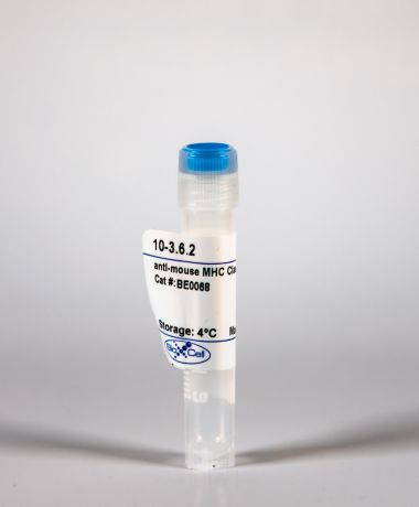
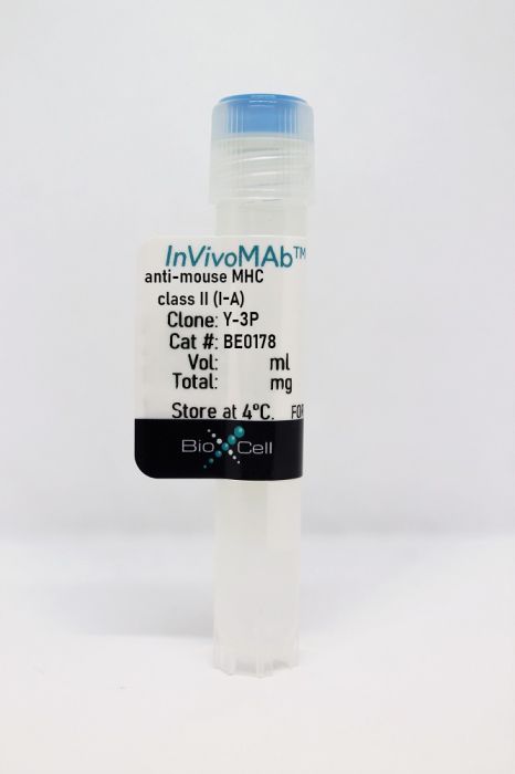
InVivoMAb anti-mouse MHC class II (I-A)
Product Details
The Y-3P monoclonal antibody reacts with mouse MHC Class II haplotypes I-Ab, I-Af, I-Ap, I-Aq, I-Ar, I-As, I-Au, I-Av, and weakly with I-Ak. The Y-3P antibody is reported to inhibit I-A-restricted T cell responses.Specifications
| Isotype | Mouse IgG2a, κ |
|---|---|
| Recommended Isotype Control(s) | InVivoMAb mouse IgG2a isotype control, unknown specificity |
| Recommended Dilution Buffer | InVivoPure pH 7.0 Dilution Buffer |
| Conjugation | This product is unconjugated. Conjugation is available via our Antibody Conjugation Services. |
| Immunogen | BALB/c x C57BL/6 F1 mouse spleen cells |
| Reported Applications |
in vivo blockade of TCR stimulation Flow cytometry |
| Formulation |
PBS, pH 7.0 Contains no stabilizers or preservatives |
| Endotoxin |
<2EU/mg (<0.002EU/μg) Determined by LAL gel clotting assay |
| Purity |
>95% Determined by SDS-PAGE |
| Sterility | 0.2 µm filtration |
| Production | Purified from cell culture supernatant in an animal-free facility |
| Purification | Protein G |
| RRID | AB_10949066 |
| Molecular Weight | 150 kDa |
| Storage | The antibody solution should be stored at the stock concentration at 4°C. Do not freeze. |
Recommended Products
-
Recommended Isotype Control(s)
InVivoMAb mouse IgG2a isotype control, unknown specificity
-

Recommended Dilution Buffer
InVivoPure pH 7.0 Dilution Buffer
in vivo blockade of TCR stimulation
Campisi, L., et al. (2016). "Apoptosis in response to microbial infection induces autoreactive TH17 cells" Nat Immunol. doi : 10.1038/ni.3512. PubMed
Microbial infections often precede the onset of autoimmunity. How infections trigger autoimmunity remains poorly understood. We investigated the possibility that infection might create conditions that allow the stimulatory presentation of self peptides themselves and that this might suffice to elicit autoreactive T cell responses that lead to autoimmunity. Self-reactive CD4+ T cells are major drivers of autoimmune disease, but their activation is normally prevented through regulatory mechanisms that limit the immunostimulatory presentation of self antigens. Here we found that the apoptosis of infected host cells enabled the presentation of self antigens by major histocompatibility complex class II molecules in an inflammatory context. This was sufficient for the generation of an autoreactive TH17 subset of helper T cells, prominently associated with autoimmune disease. Once induced, the self-reactive TH17 cells promoted auto-inflammation and autoantibody generation. Our findings have implications for how infections precipitate autoimmunity.
in vivo blockade of TCR stimulation
Feng, Y., et al. (2015). "A mechanism for expansion of regulatory T-cell repertoire and its role in self-tolerance" Nature. doi : 10.1038/nature16141. PubMed
T-cell receptor (TCR) signalling has a key role in determining T-cell fate. Precursor cells expressing TCRs within a certain low-affinity range for complexes of self-peptide and major histocompatibility complex (MHC) undergo positive selection and differentiate into naive T cells expressing a highly diverse self-MHC-restricted TCR repertoire. In contrast, precursors displaying TCRs with a high affinity for ‘self’ are either eliminated through TCR-agonist-induced apoptosis (negative selection) or restrained by regulatory T (Treg) cells, whose differentiation and function are controlled by the X-chromosome-encoded transcription factor Foxp3 (reviewed in ref. 2). Foxp3 is expressed in a fraction of self-reactive T cells that escape negative selection in response to agonist-driven TCR signals combined with interleukin 2 (IL-2) receptor signalling. In addition to Treg cells, TCR-agonist-driven selection results in the generation of several other specialized T-cell lineages such as natural killer T cells and innate mucosal-associated invariant T cells. Although the latter exhibit a restricted TCR repertoire, Treg cells display a highly diverse collection of TCRs. Here we explore in mice whether a specialized mechanism enables agonist-driven selection of Treg cells with a diverse TCR repertoire, and the importance this holds for self-tolerance. We show that the intronic Foxp3 enhancer conserved noncoding sequence 3 (CNS3) acts as an epigenetic switch that confers a poised state to the Foxp3 promoter in precursor cells to make Treg cell lineage commitment responsive to a broad range of TCR stimuli, particularly to suboptimal ones. CNS3-dependent expansion of the TCR repertoire enables Treg cells to control self-reactive T cells effectively, especially when thymic negative selection is genetically impaired. Our findings highlight the complementary roles of these two main mechanisms of self-tolerance.
in vivo blockade of TCR stimulation
Guo, L., et al. (2015). "Innate immunological function of TH2 cells in vivo" Nat Immunol 16(10): 1051-1059. PubMed
Type 2 helper T cells (TH2 cells) produce interleukin 13 (IL-13) when stimulated by papain or house dust mite extract (HDM) and induce eosinophilic inflammation. This innate response is dependent on IL-33 but not T cell antigen receptors (TCRs). While type 2 innate lymphoid cells (ILC2 cells) are the dominant innate producers of IL-13 in naive mice, we found here that helminth-infected mice had more TH2 cells compared to uninfected mice, and thes e cells became major mediators of innate type 2 responses. TH2 cells made important contributions to HDM-induced antigen-nonspecific eosinophilic inflammation and protected mice recovering from infection with Ascaris suum against subsequent infection with the phylogenetically distant nematode Nippostrongylus brasiliensis. Our findings reveal a previously unappreciated role for effector TH2 cells during TCR-independent innate-like immune responses.
in vivo blockade of TCR stimulation
Younes, S. A., et al. (2011). "Memory phenotype CD4 T cells undergoing rapid, nonburst-like, cytokine-driven proliferation can be distinguished from antigen-experienced memory cells" PLoS Biol 9(10): e1001171. PubMed
Memory phenotype (CD44(bright), CD25(negative)) CD4 spleen and lymph node T cells (MP cells) proliferate rapidly in normal or germ-free donors, with BrdU uptake rates of 6% to 10% per day and Ki-67 positivity of 18% to 35%. The rapid proliferation of MP cells stands in contrast to the much slower proliferation of lymphocytic choriomeningitis virus (LCMV)-specific memory cells that divide at rates ranging from <1% to 2% per day over the period from 15 to 60 days after LCMV infection. Anti-MHC class II antibodies fail to inhibit the in situ proliferation of MP cells, implying a non-T-cell receptor (TCR)-driven proliferation. Such proliferation is partially inhibited by anti-IL-7Ralpha antibody. The sequence diversity of TCRbeta CDR3 gene segments is comparable among the proliferating and quiescent MP cells from conventional and germ-free mice, implying that the majority of proliferating MP cells have not recently derived from a small cohort of cells that expand through multiple continuous rounds of cell division. We propose that MP cells constitute a diverse cell population, containing a subpopulation of slowly dividing authentic antigen-primed memory cells and a majority population of rapidly proliferating cells that did not arise from naive cells through conventional antigen-driven clonal expansion.
Flow Cytometry
Wei, J., et al. (2011). "Tissue-specific expression of B7x protects from CD4 T cell-mediated autoimmunity" J Exp Med 208(8): 1683-1694. PubMed
B7x, an inhibitory member of the B7/CD28 superfamily, is highly expressed in a broad range of nonhematopoietic organs, suggesting a role in maintaining peripheral tolerance. As endogenous B7x protein is expressed in pancreatic islets, we investigated whether the molecule inhibits diabetogenic responses. Transfer of disease-inducing BDC2.5 T cells into B7x-deficient mice resulted in a more aggressive form of diabetes than in wild-type animals. This exacerbation of disease correlated with higher frequencies of islet-infiltrating Th1 and Th17 cells. Conversely, local B7x overexpression inhibited the development of autoimmunity, as crossing diabetes-susceptible BDC2.5/B6(g7) mice to animals overexpressing B7x in pancreatic islets abrogated disease induction. This protection was caused by the inhibition of IFN-gamma production by CD4 T cells and not to a skewing or expansion of Th2 or regulatory T cells. The suppressive function of B7x was also supported by observations from another autoimmune model, experimental autoimmune encephalomyelitis, in which B7x-deficient mice developed exacerbated disease in comparison with wild-type animals. Analysis of central nervous system-infiltrating immune cells revealed that the loss of endogenous B7x resulted in expanded Th1 and Th17 responses. Data from these two autoimmune models provide evidence that B7x expression in the periphery acts as an immune checkpoint to prevent tissue-specific autoimmunity.
in vivo blockade of TCR stimulation
Andersson, J., et al. (2007). "CD4+CD25+ regulatory T cells are activated in vivo by recognition of self" Int Immunol 19(4): 557-566. PubMed
Naturally occurring CD4(+)CD25(+) regulatory T cells (nT(R)) comprise a separate lineage of T cells that are essential for maintaining immunological tolerance to self. Here we demonstrate that the level of phosphorylation of the TCR zeta-chain is approximately 1.5- to 4-fold higher in nT(R) as compared with CD4(+)CD25(-) T cells. The increased level of TCR zeta-chain phosphorylation is presumably secondary to their higher affinity for self, resulting in a stronger TCR signal as it was completely blocked by treatment with anti-MHC class II. The enhanced level of TCR zeta-chain phosphorylation was correlated with the capacity of nT(R) to develop non-specific suppressor effector function following culture with IL-2 or IL-4 in the absence of TCR stimulus. Thus, a sub-population of nT(R) is activated by recognition of self-peptide-MHC class II ligands in vivo, resulting in their capacity to be induced to mediate suppressor function in vitro in the absence of TCR stimulation.