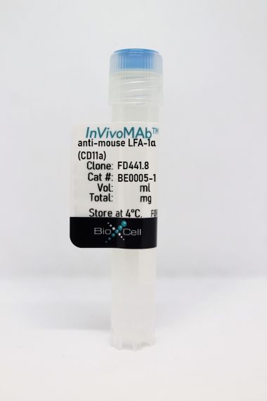
InVivoMAb anti-mouse LFA-1α (CD11a)

Product Details
The M17/4 monoclonal antibody reacts with mouse LFA-1α (lymphocyte function-associated antigen 1 alpha) also known as integrin alpha L chain and CD11a. LFA-1α and CD18 combine to form LFA-1, a 180 kDa glycoprotein and a member of the integrin family. LFA-1 is expressed on the surface of all leukocytes including lymphocytes, monocytes, macrophages, and granulocytes. LFA-1 plays a central role in leukocyte intercellular adhesion through interactions with its ligands, ICAM-1 (CD54), ICAM-2 (CD102), and ICAM-3 (CD50), and also functions in lymphocyte costimulatory signaling.Specifications
| Isotype | Rat IgG2a, κ |
|---|---|
| Recommended Isotype Control(s) | InVivoMAb rat IgG2a isotype control, anti-trinitrophenol |
| Recommended Dilution Buffer | InVivoPure pH 7.0 Dilution Buffer |
| Conjugation | This product is unconjugated. Conjugation is available via our Antibody Conjugation Services. |
| Immunogen | C57BL/6 mouse splenic secondary cytotoxic T cells |
| Reported Applications |
in vivo LFA-1 neutralization Flow cytometry |
| Formulation |
PBS, pH 7.0 Contains no stabilizers or preservatives |
| Endotoxin |
<2EU/mg (<0.002EU/μg) Determined by LAL gel clotting assay |
| Purity |
>95% Determined by SDS-PAGE |
| Sterility | 0.2 µm filtration |
| Production | Purified from cell culture supernatant in an animal-free facility |
| Purification | Protein G |
| RRID | AB_1107578 |
| Molecular Weight | 150 kDa |
| Storage | The antibody solution should be stored at the stock concentration at 4°C. Do not freeze. |
Recommended Products
-
Recommended Isotype Control(s)
InVivoMAb rat IgG2a isotype control, anti-trinitrophenol
-

Recommended Dilution Buffer
InVivoPure pH 7.0 Dilution Buffer
in vivo LFA-1 neutralization
Peske, J. D., et al. (2015). "Effector lymphocyte-induced lymph node-like vasculature enables naive T-cell entry into tumours and enhanced anti-tumour immunity" Nat Commun 6: 7114. PubMed
The presence of lymph node (LN)-like vasculature in tumours, characterized by expression of peripheral node addressin and chemokine CCL21, is correlated with T-cell infiltration and positive prognosis in breast cancer and melanoma patients. However, mechanisms controlling the development of LN-like vasculature and how it might contribute to a beneficial outcome for cancer patients are unknown. Here we demonstrate that LN-like vasculature is present in murine models of melanoma and lung carcinoma. It enables infiltration by naive T cells that significantly delay tumour outgrowth after intratumoral activation. Development of this vasculature is controlled by a mechanism involving effector CD8 T cells and NK cells that secrete LTalpha3 and IFNgamma. LN-like vasculature is also associated with organized aggregates of B lymphocytes and gp38(+) fibroblasts, which resemble tertiary lymphoid organs that develop in models of chronic inflammation. These results establish LN-like vasculature as both a consequence of and key contributor to anti-tumour immunity.
in vivo LFA-1 neutralization
Guidotti, L. G., et al. (2015). "Immunosurveillance of the liver by intravascular effector CD8(+) T cells" Cell 161(3): 486-500. PubMed
Effector CD8(+) T cells (CD8 TE) play a key role during hepatotropic viral infections. Here, we used advanced imaging in mouse models of hepatitis B virus (HBV) pathogenesis to understand the mechanisms whereby these cells home to the liver, recognize antigens, and deploy effector functions. We show that circulating CD8 TE arrest within liver sinusoids by docking onto platelets previously adhered to sinusoidal hyaluronan via CD44. After the initial arrest, CD8 TE actively crawl along liver sinusoids and probe sub-sinusoidal hepatocytes for the presence of antigens by extending cytoplasmic protrusions through endothelial fenestrae. Hepatocellular antigen recognition triggers effector functions in a diapedesis-independent manner and is inhibited by the processes of sinusoidal defenestration and capillarization that characterize liver fibrosis. These findings reveal the dynamic behavior whereby CD8 TE control hepatotropic pathogens and suggest how liver fibrosis might reduce CD8 TE immune surveillance toward infected or transformed hepatocytes.
in vivo LFA-1 neutralization, Flow Cytometry
Glatigny, S., et al. (2015). "Integrin alpha L controls the homing of regulatory T cells during CNS autoimmunity in the absence of integrin alpha 4" Sci Rep 5: 7834. PubMed
Experimental autoimmune encephalomyelitis (EAE), the animal model of multiple sclerosis (MS), results from an autoimmune attack of the central nervous system (CNS) by effector T helper (Th) 1 and Th17 cells. Regulatory T cells (Treg) can control effector T cells and limit the progression of CNS autoimmunity. Integrin alpha 4 (Itga4) is critical for the entry of Th1 but not Th17 cells into the CNS during EAE. Whether Itga4 controls the homing of Tregs in the CNS and whether Tregs can limit Th17-mediated EAE has, however, not been addressed. Through selective elimination of Itga4 in Foxp3-expressing cells, we show here that Tregs can suppress Th17-mediated EAE and enter into the CNS independently of Itga4. Furthermore, similarly to Th17 cells and in contrast to Th1 cells, Tregs depend on LFA-1 for their entry into the CNS in the absence of Itga4. Therefore, these data suggest that the efficacy of Itga4 neutralization on MS progression may be associated with the prevention of Th1 cells and the maintenance of Tregs migration into the CNS.
in vivo LFA-1 neutralization
Zenaro, E., et al. (2015). "Neutrophils promote Alzheimer’s disease-like pathology and cognitive decline via LFA-1 integrin" Nat Med 21(8): 880-886. PubMed
Inflammation is a pathological hallmark of Alzheimer’s disease, and innate immune cells have been shown to contribute to disease pathogenesis. In two transgenic models of Alzheimer’s disease (5xFAD and 3xTg-AD mice), neutrophils extravasated and were present in areas with amyloid-beta (Abeta) deposits, where they released neutrophil extracellular traps (NETs) and IL-17. Abeta42 peptide triggered the LFA-1 integrin high-affinity state and rapid neutrophil adhesion to integrin ligands. In vivo, LFA-1 integrin controlled neutrophil extravasation into the CNS and intraparenchymal motility. In transgenic Alzheimer’s disease models, neutrophil depletion or inhibition of neutrophil trafficking via LFA-1 blockade reduced Alzheimer’s disease-like neuropathology and improved memory in mice already showing cognitive dysfunction. Temporary depletion of neutrophils for 1 month at early stages of disease led to sustained improvements in memory. Transgenic Alzheimer’s disease model mice lacking LFA-1 were protected from cognitive decline and had reduced gliosis. In humans with Alzheimer’s disease, neutrophils adhered to and spread inside brain venules and were present in the parenchyma, along with NETs. Our results demonstrate that neutrophils contribute to Alzheimer’s disease pathogenesis and cognitive impairment and suggest that the inhibition of neutrophil trafficking may be beneficial in Alzheimer’s disease.
in vivo LFA-1 neutralization
Rabenstein, H., et al. (2014). "Differential kinetics of antigen dependency of CD4+ and CD8+ T cells" J Immunol 192(8): 3507-3517. PubMed
Ag recognition via the TCR is necessary for the expansion of specific T cells that then contribute to adaptive immunity as effector and memory cells. Because CD4+ and CD8+ T cells differ in terms of their priming APCs and MHC ligands we compared their requirements of Ag persistence during their expansion phase side by side. Proliferation and effector differentiation of TCR transgenic and polyclonal mouse T cells were thus analyzed after transient and continuous TCR signals. Following equally strong stimulation, CD4+ T cell proliferation depended on prolonged Ag presence, whereas CD8+ T cells were able to divide and differentiate into effector cells despite discontinued Ag presentation. CD4+ T cell proliferation was neither affected by Th lineage or memory differentiation nor blocked by coinhibitory signals or missing inflammatory stimuli. Continued CD8+ T cell proliferation was truly independent of self-peptide/MHC-derived signals. The subset divergence was also illustrated by surprisingly broad transcriptional differences supporting a stronger propensity of CD8+ T cells to programmed expansion. These T cell data indicate an intrinsic difference between CD4+ and CD8+ T cells regarding the processing of TCR signals for proliferation. We also found that the presentation of a MHC class II-restricted peptide is more efficiently prolonged by dendritic cell activation in vivo than a class I bound one. In summary, our data demonstrate that CD4+ T cells require continuous stimulation for clonal expansion, whereas CD8+ T cells can divide following a much shorter TCR signal.
in vivo LFA-1 neutralization
Hock, K., et al. (2014). "Donor CD4 T cells trigger costimulation blockade-resistant donor bone marrow rejection through bystander activation requiring IL-6" Am J Transplant 14(9): 2011-2022. PubMed
Bone marrow (BM) transplantation under costimulation blockade induces chimerism and tolerance. Cotransplantation of donor T cells (contained in substantial numbers in mobilized peripheral blood stem cells and donor lymphocyte infusions) together with donor BM paradoxically triggers rejection of donor BM through undefined mechanisms. Here, nonmyeloablatively irradiated C57BL/6 recipients simultaneously received donor BM (BALB/c) and donor T cells under costimulation blockade (anti-CD154 and CTLA4Ig). Donor CD4, but not CD8 cells, triggered natural killer-independent donor BM rejection which was associated with increased production of IL-6, interferon gamma (IFN-gamma) and IL-17A. BM rejection was prevented through neutralization of IL-6, but not of IFN-gamma or IL-17A. IL-6 counteracted the antiproliferative effect of anti-CD154 in vitro. Rapamycin and anti-lymphocyte function-associated antigen 1 negated this effect of IL-6 in vitro and prevented BM rejection in vivo. Simultaneous cotransplantation of (BALB/cxB6)F1, recipient or irradiated donor CD4 cells, or late transfer of donor CD4 cells did not lead to BM rejection, whereas cotransplantation of third party CD4 cells did. Transferred donor CD4 cells became activated, rapidly underwent apoptosis and triggered activation and proliferation of recipient T cells. Collectively, these results provide evidence that donor T cells recognizing the recipient as allogeneic lead to the release of IL-6, which abolishes the effect of anti-CD154, triggering donor BM rejection through bystander activation.
Flow Cytometry
Wang, X., et al. (2014). "Integrin-mediated interactions between B cells and follicular dendritic cells influence germinal center B cell fitness" J Immunol 192(10): 4601-4609. PubMed
Integrin-ligand interactions between germinal center (GC) B cells and Ag-presenting follicular dendritic cells (FDCs) have been suggested to play central roles during GC responses, but their in vivo requirement has not been directly tested. In this study, we show that, whereas integrins alphaLbeta2 and alpha4beta1 are highly expressed and functional on mouse GC B cells, removal of single integrins or their ligands had little effect on B cell participation in the GC response. Combined beta2 integrin deficiency and alpha4 integrin blockade also did not affect the GC response against a particulate Ag. However, the combined integrin deficiency did cause B cells to be outcompeted in splenic GC responses against a soluble protein Ag and in mesenteric lymph node GC responses against gut-derived Ags. Similar findings were made for beta2-deficient B cells in mice lacking VCAM1 on FDCs. The reduced fitness of the GC B cells did not appear to be due to decreased Ag acquisition, proliferation rates, or pAKT levels. In summary, our findings provide evidence that alphaLbeta2 and alpha4beta1 play overlapping and context-dependent roles in supporting interactions with FDCs that can augment the fitness of responding GC B cells. We also find that mouse GC B cells upregulate alphavbeta3 and adhere to vitronectin and milk-fat globule epidermal growth factor VIII protein. Integrin beta3-deficient B cells contributed in a slightly exaggerated manner to GC responses, suggesting this integrin has a regulatory function in GC B cells.
in vivo LFA-1 neutralization
Gerard, A., et al. (2013). "Secondary T cell-T cell synaptic interactions drive the differentiation of protective CD8+ T cells" Nat Immunol 14(4): 356-363. PubMed
Immunization results in the differentiation of CD8+ T cells, such that they acquire effector abilities and convert into a memory pool. Priming of T cells takes place via an immunological synapse formed with an antigen-presenting cell (APC). By disrupting synaptic stability at different times, we found that the differentiation of CD8+ T cells required cell interactions beyond those made with APCs. We identified a critical differentiation period that required interactions between primed T cells. We found that T cell-T cell synapses had a major role in the generation of protective CD8+ T cell memory. T cell-T cell synapses allowed T cells to polarize critical secretion of interferon-gamma (IFN-gamma) toward each other. Collective activation and homotypic clustering drove cytokine sharing and acted as regulatory stimuli for T cell differentiation.
in vivo LFA-1 neutralization
Li, W., et al. (2012). "Intravital 2-photon imaging of leukocyte trafficking in beating heart" J Clin Invest 122(7): 2499-2508. PubMed
Two-photon intravital microscopy has substantially broadened our understanding of tissue- and organ-specific differences in the regulation of inflammatory responses. However, little is known about the dynamic regulation of leukocyte recruitment into inflamed heart tissue, largely due to technical difficulties inherent in imaging moving tissue. Here, we report a method for imaging beating murine hearts using intravital 2-photon microscopy. Using this method, we visualized neutrophil trafficking at baseline and during inflammation. Ischemia reperfusion injury induced by transplantation or transient coronary artery ligation led to recruitment of neutrophils to the heart, their extravasation from coronary veins, and infiltration of the myocardium where they formed large clusters. Grafting hearts containing mutant ICAM-1, a ligand important for neutrophil recruitment, reduced the crawling velocities of neutrophils within vessels, and markedly inhibited their extravasation. Similar impairment was seen with the inhibition of Mac-1, a receptor for ICAM-1. Blockade of LFA-1, another ICAM-1 receptor, prevented neutrophil adherence to endothelium and extravasation in heart grafts. As inflammatory responses in the heart are of great relevance to public health, this imaging approach holds promise for studying cardiac-specific mechanisms of leukocyte recruitment and identifying novel therapeutic targets for treating heart disease.
in vivo LFA-1 neutralization
Thomas, S. Y., et al. (2011). "PLZF induces an intravascular surveillance program mediated by long-lived LFA-1-ICAM-1 interactions" J Exp Med 208(6): 1179-1188. PubMed
Innate-like NKT cells conspicuously accumulate within the liver microvasculature of healthy mice, crawling on the luminal side of endothelial cells, but their general recirculation pattern and the mechanism of their intravascular behavior have not been elucidated. Using parabiotic mice, we demonstrated that, despite their intravascular location, most liver NKT cells failed to recirculate. Antibody blocking experiments established that they were retained locally through constitutive LFA-1-intercellular adhesion molecule (ICAM) 1 interactions. This unprecedented lifelong intravascular residence could be induced in conventional CD4 T cells by the sole expression of promyelocytic leukemia zinc finger (PLZF), a transcription factor specifically expressed in the NKT lineage. These findings reveal the unique genetic and biochemical pathway that underlies the innate intravascular surveillance program of NKT cells.
in vivo LFA-1 neutralization
Pearl, J. I., et al. (2011). "Short-term immunosuppression promotes engraftment of embryonic and induced pluripotent stem cells" Cell Stem Cell 8(3): 309-317. PubMed
Embryonic stem cells (ESCs) are an attractive source for tissue regeneration and repair therapies because they can be differentiated into virtually any cell type in the adult body. However, for this approach to succeed, the transplanted ESCs must survive long enough to generate a therapeutic benefit. A major obstacle facing the engraftment of ESCs is transplant rejection by the immune system. Here we show that blocking leukocyte costimulatory molecules permits ESC engraftment. We demonstrate the success of this immunosuppressive therapy for mouse ESCs, human ESCs, mouse induced pluripotent stem cells (iPSCs), human induced pluripotent stem cells, and more differentiated ESC/(iPSCs) derivatives. Additionally, we provide evidence describing the mechanism by which inhibition of costimulatory molecules suppresses T cell activation. This report describes a short-term immunosuppressive approach capable of inducing engraftment of transplanted ESCs and iPSCs, providing a significant improvement in our mechanistic understanding of the critical role costimulatory molecules play in leukocyte activation.
in vivo LFA-1 neutralization, Flow Cytometry
Rothhammer, V., et al. (2011). "Th17 lymphocytes traffic to the central nervous system independently of alpha4 integrin expression during EAE" J Exp Med 208(12): 2465-2476. PubMed
The integrin alpha4beta1 (VLA-4) is used by encephalitogenic T cells to enter the central nervous system (CNS). However, both Th1 and Th17 cells are capable of inducing experimental autoimmune encephalomyelitis (EAE), and the molecular cues mediating the infiltration of Th1 versus Th17 cells into the CNS have not yet been defined. We investigated how blocking of alpha4 integrins affected trafficking of Th1 and Th17 cells into the CNS during EAE. Although antibody-mediated inhibition of alpha4 integrins prevented EAE when MOG(35-55)-specific Th1 cells were adoptively transferred, Th17 cells entered the brain, but not the spinal cord parenchyma, irrespective of alpha4 blockade. Accordingly, T cell-conditional alpha4-deficient mice were not resistant to actively induced EAE but showed an ataxic syndrome with predominantly supraspinal infiltrates of IL-23R(+)CCR6(+)CD4(+) T cells. The entry of alpha4-deficient Th17 cells into the CNS was abolished by blockade of LFA-1 (alphaLbeta2 integrin). Thus, Th1 cells preferentially infiltrate the spinal cord via an alpha4 integrin-mediated mechanism, whereas the entry of Th17 cells into the brain parenchyma occurs in the absence of alpha4 integrins but is dependent on the expression of alphaLbeta2. These observations have implications for the understanding of lesion localization, immunosurveillance, and drug design in multiple sclerosis.