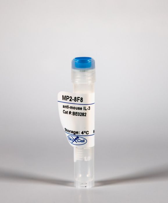
InVivoMab anti-mouse IL-3
Product Details
The MP2-8F8 monoclonal antibody reacts with mouse IL-3, a pleiotropic cytokine produced primarily by activated T cells but also by activated mast cells, keratinocytes and neurons/astrocytes. IL-3 is highly species-specific, mature human and mouse IL-3 share only 29% sequence identity and human IL-3 does not show activity on mouse cells. IL-3 can stimulate the proliferation and differentiation of megakaryocytes, neutrophils, and macrophages from bone marrow cultures. Additionally, IL-3 also affects the functional activity of mature mast cells, basophils, eosinophils and macrophages. The MP2-8F8 antibody has been reported to neutralize the bioactivity of natural or recombinant IL-3 as well as stimulate basophil proliferation when administered as a complex with IL-3.Specifications
| Isotype | Rat IgG1 |
|---|---|
| Recommended Isotype Control(s) | InVivoMAb rat IgG1 isotype control, anti-horseradish peroxidase |
| Recommended Dilution Buffer | InVivoPure pH 6.5 Dilution Buffer |
| Conjugation | This product is unconjugated. Conjugation is available via our Antibody Conjugation Services. |
| Immunogen | COS-expressed, recombinant mouse IL-3 |
| Reported Applications |
in vivo IL-3 neutralization in vitro IL-3 neutralization in vivo IL-3 receptor stimulation (as a complex with IL-3) ELISA Flow cytometry |
| Formulation |
PBS, pH 6.5 Contains no stabilizers or preservatives |
| Endotoxin |
<2EU/mg (<0.002EU/μg) Determined by LAL gel clotting assay |
| Purity |
>95% Determined by SDS-PAGE |
| Sterility | 0.2 μM filtered |
| Production | Purified from cell culture supernatant in an animal-free facility |
| Purification | Protein G |
| RRID | AB_2687805 |
| Molecular Weight | 150 kDa |
| Storage | The antibody solution should be stored at the stock concentration at 4°C. Do not freeze. |
Recommended Products
-
Recommended Isotype Control(s)
InVivoMAb rat IgG1 isotype control, anti-horseradish peroxidase
-
Recommended Dilution Buffer
InVivoPure pH 6.5 Dilution Buffer
in vivo IL-3 receptor stimulation (as a complex with IL-3)
Kamran, N., et al. (2018). "Melanoma induced immunosuppression is mediated by hematopoietic dysregulation" Oncoimmunology 7(3): e1408750. PubMed
Tumors are associated with expansion of immunosuppressive cells such as tumor associated macrophages (TAMs), regulatory T cells (Tregs) and myeloid derived suppressor cells (MDSCs). These cells promote tumor growth, angiogenesis, metastasis and immune escape. Cancer patients frequently present symptoms such as anemia, leukocytosis and/or cytopenia; associated with poor prognosis. To uncover tumor-mediated hematopoietic abnormalities and identify novel targets that can be harnessed to improve tumor-specific immune responses, we investigated the hematopoietic stem and progenitor cell compartment in melanoma bearing mice. We show that melanoma growth results in expansion of myeloid lineages such as MDSCs, macrophages and DCs along with a reduction in mature RBCs and platelets. Mature B lymphocytes in the blood and BM of melanoma mice were also reduced. Mice bearing melanoma showed extramedullary hematopoiesis in the spleen. Increased expansion of myeloid lineages occurred directly at the level of stem and progenitor cells. The reduction in mature B lymphocytes resulted from a block at the Pro-B cell stage in the bone marrow. Addition of recombinant IL-3 to bone marrow cells resulted in the expansion of committed myeloid progenitors including common myeloid precursors, granulocyte-monocyte precursors and megakaryocyte-erythrocyte precursors. In vivo, IL-3 receptor stimulation in melanoma bearing mice using an IL-3 antibody also resulted in a robust expansion of committed myeloid progenitors and hematopoietic stem cells. Collectively our findings demonstrate that tumor growth plays a pivotal role in reprogramming the host immune system by impacting hematopoiesis directly at the level of stem cell compartment.
in vivo IL-3 neutralization
Renner, K., et al. (2015). "IL-3 contributes to development of lupus nephritis in MRL/lpr mice" Kidney Int 88(5): 1088-1098. PubMed
MRL/lpr mice develop a spontaneous autoimmune disease that closely resembles human systemic lupus erythematosus (SLE) with DNA autoantibodies, hypergammaglobulinemia, immune complex glomerulonephritis, and systemic vasculitis. Little is known about the role of IL-3 in SLE. In order to study this we analyzed the expression of IL-3 in murine lupus and determined whether blockade of IL-3 with a monoclonal antibody or injection of recombinant IL-3 affects lupus nephritis in MRL/lpr mice. During disease progression IL-3 levels were increased in the plasma and in the supernatant of cultured splenocytes from MRL/lpr mice. Administration of IL-3 aggravated the disease with significantly higher renal activity scores, more renal fibrosis, and more glomerular leukocyte infiltration and IgG deposition. Blockade of IL-3 significantly improved acute and chronic kidney damage, reduced the glomerular infiltration of leukocytes and the glomerular deposition of IgG, and decreased the development of renal fibrosis. Furthermore, DNA autoantibody production, proteinuria, and serum creatinine levels were significantly lower in the anti-IL-3 group. Thus, IL-3 plays an important role in the pathogenesis of SLE and the progression of lupus nephritis. Hence, blockade of IL-3 may represent a new strategy for treatment of lupus nephritis.
in vivo IL-3 receptor stimulation (as a complex with IL-3)
Li, Y., et al. (2015). "The STAT5-GATA2 pathway is critical in basophil and mast cell differentiation and maintenance" J Immunol 194(9): 4328-4338. PubMed
Transcription factor GATA binding protein 2 (GATA2) plays critical roles in hematopoietic stem cell survival and proliferation, granulocyte-monocyte progenitor differentiation, and basophil and mast cell differentiation. However, precise roles of GATA2 in basophil and mast cell differentiation and maintenance have not been delineated. We have identified GATA2 as an essential transcription factor in differentiation of newly identified common basophil and mast cell progenitors into basophils and mast cells. We observed Gata2 haploinsufficiency for mast cell differentiation, but not for basophil differentiation. We examined the precise role of GATA2 in maintaining the expression of a wide range of genes that are important for performing basophil or mast cell functions. The effects of GATA2 on gene expression were broadly based. We demonstrated that GATA2 was required for maintaining Fcer1a mRNA and FcepsilonRIalpha protein expression on both basophils and mast cells, as well as for maintaining Kit mRNA and c-Kit protein expression on mast cells. GATA2 was required for histamine synthesis and was also critical for Il4 mRNA expression in basophils and Il13 mRNA expression in mast cells. We demonstrate a STAT5-GATA2 connection, showing that the STAT5 transcription factor directly bound to the promoter and an intronic region of the Gata2 gene. Overexpression of the Gata2 gene was sufficient to direct basophil and mast cell differentiation in the absence of the Stat5 gene. Our study reveals that the STAT5-GATA2 pathway is critical for basophil and mast cell differentiation and maintenance.
in vivo IL-3 receptor stimulation (as a complex with IL-3)
Shiraishi, Y., et al. (2013). "Sequential engagement of FcepsilonRI on Mast Cells and Basophil Histamine H(4) Receptor and FcepsilonRI in Allergic Rhinitis" J Immunol 190(2): 539-548. PubMed
Histamine H(4) receptor (H(4)R)-deficient mice (H(4)R(-/-)), H(4)R antagonist-treated wild-type (WT) mice, and WT mice depleted of basophils failed to develop early (EPR) or late phase (LPR) nasal responses following allergen sensitization and challenge. Basophil transfer from WT but not H(4)R(-/-) mice restored the EPR and LPR in H(4)R(-/-) mice. Following passive sensitization with OVA-specific IgE, FcepsilonRI(-/-) recipients of WT basophils plus OVA and histamine developed an EPR and LPR. OVA-IgE passively sensitized FcepsilonRI(-/-) recipients of H(4)R(-/-) basophils and OVA and histamine challenge failed to develop an EPR or LPR, and basophils were not detected in nasal tissue. In contrast, recipients of basophils from IL-13(-/-) and IL-4(-/-)/IL-13(-/-) mice developed an EPR but not an LPR. These results demonstrate the development of allergic rhinitis proceeded in two distinct stages: histamine release from FcepsilonRI-activated mast cells, followed by histamine-mediated recruitment of H(4)R-expressing basophils to the nasal cavity and activation through FcepsilonRI.
Flow Cytometry
Leyva-Castillo, J. M., et al. (2013). "Skin thymic stromal lymphopoietin initiates Th2 responses through an orchestrated immune cascade" Nat Commun 4: 2847. PubMed
Thymic stromal lymphopoietin (TSLP) has emerged as a key initiator in Th2 immune responses, but the TSLP-driven immune cascade leading to Th2 initiation remains to be delineated. Here, by dissecting the cellular network triggered by mouse skin TSLP in vivo, we uncover that TSLP-promoted IL-4 induction in CD4(+) T cells in skin-draining lymph nodes is driven by an orchestrated ‘DC-T-Baso-T’ cascade, which represents a sequential cooperation of dendritic cells (DCs), CD4(+) T cells and basophils. Moreover, we reveal that TSLP-activated DCs prime naive CD4(+) T cells to produce IL-3 via OX40L signalling and demonstrate that the OX40L-IL-3 axis has a critical role in mediating basophil recruitment, CD4(+) T-cell expansion and Th2 priming. These findings thus add novel insights into the cellular network and signal axis underlying the initiation of Th2 immune responses.
ELISA
Herbst, T., et al. (2012). "Antibodies and IL-3 support helminth-induced basophil expansion" Proc Natl Acad Sci U S A 109(37): 14954-14959. PubMed
Basophils are powerful mediators of Th2 immunity and are present in increased numbers during allergic inflammation and helminth infection. Despite their ability to potentiate Th2 immunity the mechanisms regulating basophil development remain largely unknown. We have found a unique role for isotype-switched antibodies in promoting helminth-induced basophil production following infection of mice with Heligmosomoides polygyrus bakeri or Nippostrongylus brasiliensis. H. polygyrus bakeri-induced basophil expansion was found to occur within the bone marrow, and to a lesser extent the spleen, and was IL-3 dependent. IL-3 was largely produced by CD4(+)CD49b(+)NK1.1(-) effector T cells at these sites, and required the IL-4Ralpha chain. However, antibody-deficient mice exhibited defective basophil mobilization despite intact T-cell IL-3 production, and supplementation of mice with immune serum could promote basophilia independently of required IL-4Ralpha signaling. Helminth-induced eosinophilia was not affected by the deficiency in isotype-switched antibodies, suggesting a direct effect on basophils rather than through priming of Th2 responses. Although normal type 2 immunity occurred in the basopenic mice following primary infection with H. polygyrus bakeri, parasite rejection following challenge infection was impaired. These data reveal a role for isotype-switched antibodies in promoting basophil expansion and effector function following helminth infection.
in vivo IL-3 receptor stimulation (as a complex with IL-3)
Ohmori, K., et al. (2009). "IL-3 induces basophil expansion in vivo by directing granulocyte-monocyte progenitors to differentiate into basophil lineage-restricted progenitors in the bone marrow and by increasing the number of basophil/mast cell progenitors in the spleen" J Immunol 182(5): 2835-2841. PubMed
Recent work has established important roles for basophils in regulating immune responses. To exert their biological functions, basophils need to be expanded to critical numbers. However, the mechanisms underlying basophil expansion remain unclear. In this study, we established that IL-3 played an important role in the rapid and specific expansion of basophils. We found that the IL-3 complex (IL-3 plus anti-IL-3 Ab) greatly facilitated the differentiation of GMPs into basophil lineage-restricted progenitors (BaPs) but not into eosinophil lineage-restricted progenitors or mast cells in the bone marrow. We also found that the IL-3 complex treatment resulted in approximately 4-fold increase in the number of basophil/mast cell progenitors (BMCPs) in the spleen. IL-3-driven basophil expansion depended on STAT5 signaling. We showed that GMPs but not common myeloid progenitors expressed low levels of IL-3 receptor. IL-3 receptor expression was dramatically up-regulated in BaPs but not eosinophil lineage-restricted progenitors. Approximately 38% of BMCPs expressed the IL-3R alpha-chain. The up-regulated IL-3 receptor expression was not affected by IL-3 or STAT5. Our findings demonstrate that IL-3 induced specific expansion of basophils by directing GMPs to differentiate into BaPs in the bone marrow and by increasing the number of BMCPs in the spleen.
in vivo IL-3 neutralization, in vivo IL-3 receptor stimulation (as a complex with IL-3)
Munitz, A., et al. (2006). "CD48 is an allergen and IL-3-induced activation molecule on eosinophils" J Immunol 177(1): 77-83. PubMed
Eosinophils are involved in a variety of allergic, parasitic, malignant, and idiopathic disorders by releasing a variety of factors including specific granule proteins, lipid mediators, and proinflammatory and immunoregulatory cytokines and chemokines. In addition, they interact with various cell types in the inflamed tissue. Yet, the mechanism of eosinophil activation is still poorly understood. Recently, we described the expression and function of the CD2-subfamily of receptors and especially 2B4 on human eosinophils. In this study we focus on CD48, the high-affinity ligand of 2B4. CD48 is a GPI-anchored protein involved in cellular activation, costimulation, and adhesion, but has not been studied on eosinophils. We demonstrate that human eosinophils from atopic asthmatics display enhanced levels of CD48 expression and that IL-3 up-regulates CD48 expression. Furthermore, cross-linking CD48 on human eosinophils triggers release of eosinophil granule proteins. Assessment of CD48 expression in a murine model of experimental asthma revealed that CD48 is induced by allergen challenge and partially regulated by IL-3. Additionally, anti-IL-3 reduces CD48 expression and the degree of airway inflammation. Thus, CD48 is an IL-3-induced activating receptor on eosinophils, likely involved in promoting allergic inflammation.
in vivo IL-3 neutralization
Min, B., et al. (2004). "Basophils produce IL-4 and accumulate in tissues after infection with a Th2-inducing parasite" J Exp Med 200(4): 507-517. PubMed
Using mice in which the eGfp gene replaced the first exon of the Il4 gene (G4 mice), we examined production of interleukin (IL)-4 during infection by the intestinal nematode Nippostrongylus brasiliensis (Nb). Nb infection induced green fluorescent protein (GFP)pos cells that were FcepsilonRIpos, CD49bbright, c-kitneg, and Gr1neg. These cells had lobulated nuclei and granules characteristic of basophils. They were found mainly in the liver and lung, to a lesser degree in the spleen, but not in the lymph nodes. Although some liver basophils from naive mice express GFP, Nb infection enhanced GFP expression and increased the number of tissue basophils. Similar basophil GFP expression was found in infected Stat6-/- mice. Basophils did not increase in number in infected Rag2-/- mice; Rag2-/- mice reconstituted with CD4 T cells allowed significant basophil accumulation, indicating that CD4 T cells can direct both tissue migration of basophils and enhanced IL-4 production. IL-4 production was immunoglobulin independent and only partially dependent on IL-3. Thus, infection with a parasite that induces a “Th2-type response” resulted in accumulation of tissue basophils, and these cells, stimulated by a non-FcR cross-linking mechanism, are a principal source of in vivo IL-4 production.
in vitro IL-3 neutralization
Kullberg, M. C., et al. (1996). "T cell-derived IL-3 induces the production of IL-4 by non-B, non-T cells to amplify the Th2-cytokine response to a non-parasite antigen in Schistosoma mansoni-infected mice" J Immunol 156(4): 1482-1489. PubMed
We describe a novel amplification mechanism underlying the increased early IL-4 production observed in Schistosoma mansoni-infected mice in response to a non-parasite Ag, sperm whale myoglobin (SwMb). Earlier studies have shown that splenic Fc epsilon R+ non-B, non-T (NBNT) cells from schistosome-infected mice secrete IL-4 after stimulation with parasite Ag. We now demonstrate that purified NBNT cells from SwMb-immunized S. mansoni-infected mice do not respond directly to SwMb, but produce IL-4 in response to IL-3. Accordingly, we show that the early SwMb-specific IL-4 response of spleen cells (SC) from immunized infected mice is dependent on IL-3 and on CD4+ T cells. Thus, most of the early SwMb-induced IL-4 from SC of infected mice appears to be produced by NBNT cells triggered by IL-3 synthesized by SwMb-specific CD4+ T cells. IL-3-induced IL-4 production was also observed in purified NBNT cells from immunized uninfected mice, but the frequency and/or IL-4-producing capacity of splenic IL-3-responsive cells was found to be 8 to 16 times higher in immunized infected animals. IL-4 production by purified CD4+ cells from immunized infected mice was also seen after SwMb stimulation, but this response showed slower kinetics than those of total SC, was IL-3-independent, and on average threefold greater than that by CD4+ cells from immunized uninfected controls. Thus, increased SwMb-induced IL-4 production in immunized S. mansoni-infected mice results from direct synthesis by CD4+ T cells, as well as their stimulation via IL-3 of an expanded population of NBNT cells. The latter pathway may serve as an amplification loop for Th2-cytokine responses.
in vitro IL-3 neutralization
Abrams, J. S. and M. K. Pearce. (1988). "Development of rat anti-mouse interleukin 3 monoclonal antibodies which neutralize bioactivity in vitro" J Immunol 140(1): 131-137. PubMed
Several rat anti-mouse interleukin 3 (IL-3) monoclonal antibodies have been developed which inhibit the biologic activity of mouse IL-3. These antibodies were produced in rats immunized with preparations of purified, recombinant mouse IL-3, obtained from transiently transfected COS7 cell supernatant. Hybridomas secreting anti-IL-3 were selected initially either on the basis of their giving a positive signal in an indirect enzyme-linked immunosorbent assay, or for their ability to inhibit the proliferation of the IL-3-dependent mouse mast cell line, MC/9. Neutralizing rat monoclonal IgG1, IgG2a, and IgG2b antibodies have been identified; these also block IL-3-induced proliferation of the NFS-60 and IC2 cell lines. These antibodies also blocked the IL-3-induced proliferation of mouse bone marrow-derived colony-forming units-culture suggesting that the same epitopes on IL-3 influence receptor recognition for both the proliferation of factor-dependent cell lines as well as normal bone marrow cells. Fab fragments produced from certain of the IgG2a-neutralizing antibodies blocked as well as the parent IgG. Antibody cross-blocking studies identified one neutralizing antibody apparently recognizing an epitope that was spatially distinct from those recognized by the other blocking antibodies tested. The development of these neutralizing rat monoclonal antibodies to mouse IL-3 should facilitate further investigation on the role of this factor in hemopoietic regulation.