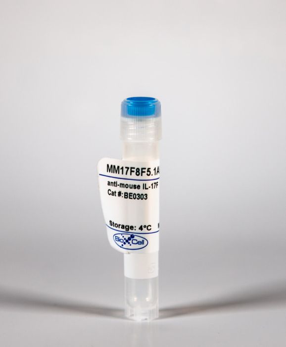
InVivoMAb anti-mouse IL-17F
Product Details
The MM17F8F5.1A9 (also known as MM17F-8F5) monoclonal antibody reacts with mouse IL-17F a 37 kDa cytokine expressed by Th17 cells, γδ T cells, mast cells, basophils, and epithelial cells. IL-17F can be secreted as homodimers or as heterodimers with IL-17A. IL -17F and IL-17A have overlapping functions. Both play an important role in anti-microbial and chronic inflammation by inducing cytokine and chemokine production, neutrophil influx, and the production of antibacterial peptides. Overexpression of IL-17F is associated with airway hyperreactivity and mucus hypersecretion. The MM17F8F5.1A9 antibody has been shown to neutralize IL-17F in vivo.Specifications
| Isotype | Mouse IgG1, κ |
|---|---|
| Recommended Isotype Control(s) | InVivoMAb mouse IgG1 isotype control, unknown specificity |
| Recommended Dilution Buffer | InVivoPure pH 7.0 Dilution Buffer |
| Conjugation | This product is unconjugated. Conjugation is available via our Antibody Conjugation Services. |
| Immunogen | Mouse IL-17F |
| Reported Applications | in vivo IL-17F neutralization |
| Formulation |
PBS, pH 7.0 Contains no stabilizers or preservatives |
| Endotoxin |
<2EU/mg (<0.002EU/μg) Determined by LAL gel clotting assay |
| Purity |
>95% Determined by SDS-PAGE |
| Sterility | 0.2 μm filtration |
| Production | Purified from cell culture supernatant in an animal-free facility |
| Purification | Protein A |
| RRID | AB_2715461 |
| Molecular Weight | 150 kDa |
| Storage | The antibody solution should be stored at the stock concentration at 4°C. Do not freeze. |
Recommended Products
-
Recommended Isotype Control(s)
InVivoMAb mouse IgG1 isotype control, unknown specificity
-

Recommended Dilution Buffer
InVivoPure pH 7.0 Dilution Buffer
in vivo IL-17F neutralization
Marchitto, M. C., et al. (2019). "Clonal Vγ6(+)Vδ4(+) T cells promote IL-17-mediated immunity against Staphylococcus aureus skin infection" Proc Natl Acad Sci U S A 116(22): 10917-10926. PubMed
T cell cytokines contribute to immunity against Staphylococcus aureus, but the predominant T cell subsets involved are unclear. In an S. aureus skin infection mouse model, we found that the IL-17 response was mediated by γδ T cells, which trafficked from lymph nodes to the infected skin to induce neutrophil recruitment, proinflammatory cytokines IL-1α, IL-1β, and TNF, and host defense peptides. RNA-seq for TRG and TRD sequences in lymph nodes and skin revealed a single clonotypic expansion of the encoded complementarity-determining region 3 amino acid sequence, which could be generated by canonical nucleotide sequences of TRGV5 or TRGV6 and TRDV4 However, only TRGV6 and TRDV4 but not TRGV5 sequences expanded. Finally, Vγ6(+) T cells were a predominant γδ T cell subset that produced IL-17A as well as IL-22, TNF, and IFNγ, indicating a broad and substantial role for clonal Vγ6(+)Vδ4(+) T cells in immunity against S. aureus skin infections.
in vivo IL-17F neutralization
Uyttenhove, C., et al. (2011). "Amine-reactive OVA multimers for auto-vaccination against cytokines and other mediators: perspectives illustrated for GCP-2 in L. major infection" J Leukoc Biol 89(6): 1001-1007. PubMed
Anticytokine auto-vaccination is a powerful tool for the study of cytokine functions in vivo but has remained rather esoteric as a result of numerous technical difficulties. We here describe a two-step procedure based on the use of OVA multimers purified by size exclusion chromatography after incubation with glutaraldehyde at pH 6. When such polymers are incubated with a target protein at pH 8.5 to deprotonate reactive amines, complexes are formed that confer immunogenicity to self-antigens. The chemokine GCP-2/CXCL6, the cytokines GM-CSF, IL-17F, IL-17E/IL-25, IL-27, and TGF-beta1, and the MMP-9/gelatinase B are discussed as examples. mAb, derived from such immunized mice, have obvious advantages for in vivo studies of the target proteins. Using a mAb against GCP-2, obtained by the method described here, we provide the first demonstration of the major role played by this chemokine in rapid neutrophil mobilization after Leishmania major infection. Pre-activated OVA multimers reactive with amine residues thus provide an efficient carrier for auto-vaccination against 9-90 kDa autologous proteins.
in vivo IL-17F neutralization
Lemaire, M. M., et al. (2011). "Dual TCR expression biases lung inflammation in DO11.10 transgenic mice and promotes neutrophilia via microbiota-induced Th17 differentiation" J Immunol 187(7): 3530-3537. PubMed
A commonly used mouse model of asthma is based on i.p. sensitization to OVA together with aluminum hydroxide (alum). In wild-type BALB/c mice, subsequent aerosol challenge using this protein generates an eosinophilic inflammation associated with Th2 cytokine expression. By constrast, in DO11.10 mice, which are transgenic for an OVA-specific TCR, the same treatment fails to induce eosinophilia, but instead promotes lung neutrophilia. In this study, we show that this neutrophilic infiltration results from increased IL-17A and IL-17F production, whereas the eosinophilic response could be restored upon blockade of IFN-gamma, independently of the Th17 response. In addition, we identified a CD4(+) cell population specifically present in DO11.10 mice that mediates the same inflammatory response upon transfer into RAG2(-/-) mice. This population contained a significant proportion of cells expressing an additional endogenous TCR alpha-chain and was not present in RAG2(-/-) DO11.10 mice, suggesting dual antigenic specificities. This particular cell population expressed markers of memory cells, secreted high levels of IL-17A, and other cytokines after short-term restimulation in vitro, and triggered a neutrophilic response in vivo upon OVA aerosol challenge. The relative numbers of these dual TCR lymphocytes increased with the age of the animals, and IL-17 production was abolished if mice were treated with large-spectrum antibiotics, suggesting that their differentiation depends on foreign Ags provided by gut microflora. Taken together, our data indicate that dual TCR expression biases the OVA-specific response in DO11.10 mice by inhibiting eosinophilic responses via IFN-gamma and promoting a neutrophilic inflammation via microbiota-induced Th17 differentiation.