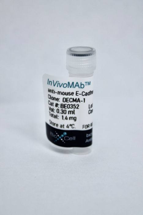
InVivoMAb anti-mouse E-Cadherin (CD324)
Product Details
The DECMA-1 monoclonal antibody reacts with the extracellular domain of mouse E-Cadherin, a 120-kDa transmembrane cell-cell adhesion glycoprotein also known as CD324 and uvomorulin. E-Cadherin is widely expressed on epithelial cells in the colon, uterus, liver, keratinocytes, brain, heart, muscle, kidney, and pancreas as well as erythroid cells. E-Cadherin is a critical regulator of epithelial junction formation. Its association with catenins is necessary for cell-to-cell adhesion. E-Cadherin is involved in development, bacterial pathogenesis, and tumor invasion. The DECMA-1 antibody has been shown to delay tumor onset and attenuate tumor burden in MMTV-PyMT mice by reducing tumor cell proliferation and inducing apoptosis without any detectable cytotoxicity.Specifications
| Isotype | Rat IgG1, κ |
|---|---|
| Recommended Isotype Control(s) | InVivoMAb rat IgG1 isotype control, anti-horseradish peroxidase |
| Recommended Dilution Buffer | InVivoPure pH 7.0 Dilution Buffer |
| Conjugation | This product is unconjugated. Conjugation is available via our Antibody Conjugation Services. |
| Immunogen | Mouse PCC4azal teratocarcinoma cells |
| Reported Applications |
in vivo E-Cadherin neutralization In vitro E-Cadherin neutralization Immunofluorescence Immunoprecipitation Western blot |
| Formulation |
PBS, pH 7.0 Contains no stabilizers or preservatives |
| Endotoxin |
<2EU/mg (<0.002EU/μg) Determined by LAL gel clotting assay |
| Purity |
>95% Determined by SDS-PAGE |
| Sterility | 0.2 μM filtered |
| Production | Purified from cell culture supernatant in an animal-free facility |
| Purification | Protein G |
| RRID | AB_2894771 |
| Molecular Weight | 150 kDa |
| Storage | The antibody solution should be stored at the stock concentration at 4°C. Do not freeze. |
Recommended Products
-
Recommended Isotype Control(s)
InVivoMAb rat IgG1 isotype control, anti-horseradish peroxidase
-

Recommended Dilution Buffer
InVivoPure pH 7.0 Dilution Buffer
Immunofluorescence
Liu, M., et al. (2020). "TGF-β suppresses type 2 immunity to cancer" Nature 587(7832): 115-120. PubMed
The immune system uses two distinct defence strategies against infections: microbe-directed pathogen destruction characterized by type 1 immunity(1), and host-directed pathogen containment exemplified by type 2 immunity in induction of tissue repair(2). Similar to infectious diseases, cancer progresses with self-propagating cancer cells inflicting host-tissue damage. The immunological mechanisms of cancer cell destruction are well defined(3-5), but whether immune-mediated cancer cell containment can be induced remains poorly understood. Here we show that depletion of transforming growth factor-β receptor 2 (TGFBR2) in CD4(+) T cells, but not CD8(+) T cells, halts cancer progression as a result of tissue healing and remodelling of the blood vasculature, causing cancer cell hypoxia and death in distant avascular regions. Notably, the host-directed protective response is dependent on the T helper 2 cytokine interleukin-4 (IL-4), but not the T helper 1 cytokine interferon-γ (IFN-γ). Thus, type 2 immunity can be mobilized as an effective tissue-level defence mechanism against cancer.
in vitro E-Cadherin neutralization, Western Blot
Xu, B., et al. (2018). "An oncolytic herpesvirus expressing E-cadherin improves survival in mouse models of glioblastoma" Nat Biotechnol . PubMed
The efficacy of oncolytic herpes simplex virus (oHSV) is limited by rapid viral clearance by innate immune effector cells and poor intratumoral viral spread. We combine two approaches to overcome these barriers: inhibition of natural killer (NK) cells and enhancement of intratumoral viral spread. We engineered an oHSV to express CDH1, encoding E-cadherin, an adherent molecule and a ligand for KLRG1, an inhibitory receptor expressed on NK cells. In vitro, infection with this engineered virus, named OV-CDH1, induced high surface E-cadherin expression on infected glioblastoma (GBM) cells, which typically lack endogenous E-cadherin. Ectopically expressed E-cadherin enhanced the spread of OV-CDH1 by facilitating cell-to-cell infection and viral entry and reduced viral clearance by selectively protecting OV-CDH1-infected cells from KLRG1(+) NK cell killing. In vivo, OV-CDH1 treatment substantially prolonged the survival in GBM-bearing mouse models, primarily because of improved viral spread rather than inhibition of NK cell activity. Thus, virus-induced overexpression of E-cadherin may be a generalizable strategy for improving cancer virotherapy.
in vivo E-Cadherin neutralization
Wang, C., et al. (2014). "Glial cell-derived neurotrophic factor attenuates neuropathic pain in a mouse model of chronic constriction injury: possible involvement of E-cadherin/p120ctn signaling" J Mol Neurosci 54(2): 156-163. PubMed
Treating neuropathic pain is a major clinical challenge, and several key molecules associated with nociception have been suggested as potential targets for novel analgesics. Many studies have reported the anti-nociceptive effects of glial cell-derived neurotrophic factor (GDNF), but the underlying mechanism remains largely unknown. The present study was performed to assess the effects of GDNF in a mouse model of chronic constriction injury (CCI)-induced neuropathic pain. We also determined the potential role of E-cadherin/p120 catenin (p120ctn) signaling in these effects. Mice received an intrathecal acute injection of PBS, GDNF, and DECMA-1 (an E-cadherin functional blocking antibody) or a combination of DECMA-1 with GDNF on the testing days. Our results demonstrated that CCI caused a rapid decrease in E-cadherin and membrane-associated p120ctn in the spinal dorsal horn. Together, these data demonstrated that E-cadherin-associated p120ctn was upregulated by GDNF and that this upregulation was inhibited by pre-treatment with DECMA-1. Moreover, DECMA-1 significantly inhibited the effect of GDNF on thermal hyperalgesia. These data suggest that GDNF might have a therapeutic potential for the treatment of CCI-induced neuropathic pain and that the E-cadherin/p120ctn might play a role in GDNF-induced attenuation of thermal hyperalgesia.
in vivo E-Cadherin neutralization, in vitro E-Cadherin neutralization, Immunofluorescence
Brouxhon, S. M., et al. (2013). "Monoclonal antibody against the ectodomain of E-cadherin (DECMA-1) suppresses breast carcinogenesis: involvement of the HER/PI3K/Akt/mTOR and IAP pathways" Clin Cancer Res 19(12): 3234-3246. PubMed
PURPOSE: Although targeted therapies against HER2 have been one of the most successful therapeutic strategies for breast cancer, patients eventually developed acquired resistance from compensatory upregulation of alternate HERs and mitogen-activated protein kinase-phosphoinositide 3-kinase (PI3K)/Akt/mTOR signaling. As we and others have shown that the soluble ectodomain fragment of E-cadherin exerts prooncogenic effects via HER1/2-mediated binding and activation of downstream prosurvival pathways, we explored whether targeting this ectodomain [DECMA-1 monoclonal antibody (mAb)] was effective in the treatment of HER2-positive (HER2(+)) breast cancers. EXPERIMENTAL DESIGN: MMTV-PyMT transgenic mice and HER2(+)/E-cadherin-positive MCF-7 and BT474 trastuzumab-resistant (TtzmR) cells were treated with the DECMA-1 mAb. Antitumor responses were assessed by bromodeoxyuridine incorporation, apoptosis, and necrosis. The underlying intracellular prooncogenic pathways were explored using subcellular fractionation, immunoprecipitation, fluorescence microscopy, and immunoblotting. RESULTS: Treatment with DECMA-1 mAb significantly delayed tumor onset and attenuated tumor burden in MMTV-PyMT mice by reducing tumor cell proliferation and inducing apoptosis without any detectable cytotoxicity to mice or end-organs. In vitro treatment of MCF-7 and BT474 TtzmR cells reduced proliferation and induced cancer cell apoptosis. Importantly, this inhibition of breast tumorigenesis was due to concomitant downregulation, via ubiquitin-mediated degradation through the lysosome and proteasome pathways, of all HER family members, components of downstream PI3K/Akt/mTOR prosurvival signaling and suppression of inhibitor of apoptosis proteins. CONCLUSIONS: Our results establish that the E-cadherin ectodomain-specific mAb DECMA-1 inhibits Ecad(+)/HER2(+) breast cancers by hindering tumor growth and inducing apoptosis via downregulation of key oncogenic pathways involved in trastuzumab resistance, thereby establishing a novel therapeutic platform for the treatment of HER2(+) breast cancers.
Immunofluorescence, Western Blot
Contreras, R. G., et al. (2002). "E-Cadherin and tight junctions between epithelial cells of different animal species" Pflugers Arch 444(4): 467-475. PubMed
The assembly and permanent sealing of tight junctions (TJs) depend crucially on cell-cell contacts containing E-cadherin. This poses a puzzling problem because, while TJs can be established between epithelial cells from different tissues and even different animal species (“heterotypic TJs”; Gonzalez-Mariscal et al. 1989, J Membr Biol 107:43), the cell-cell binding mediated by E-cadherin is a highly specific one (Takeichi 1995, Curr Opin Cell Biol 7:619). Yet the demonstration that TJs can be established at heterotypic borders is open to two distinct challenges. First, it is based on transepithelial electrical resistance (TER) and restriction to ruthenium red permeation only, which today are known to be just two of the many characteristics of TJs; and second some attributes of the TJs (e.g. the presence of specific molecules) have been found even in cells that do not establish these structures. This raised the question of whether heterotypic TJs were not true or full TJs. In the present work we demonstrate that heterotypic TJs in mixed monolayers of MDCK cells with a different cell type (LLC-PK1) are true TJs through several criteria, such as TER, the ability to stop the membrane diffusion of fluorescent sphingomyelin from the apical to the lateral domain, the presence of ZO-1, ZO-2, occludin, claudin-1 and claudin-2. We then turn to the presence of E-cadherin at heterotypic borders, and observe that it cannot be detected by the highly specific DECMA-1 antibody, in spite of the fact that this antibody does reveal the presence of E-cadherin at homotypic contacts of the same cell. Yet, ECCD-2, an antibody against another domain of E-cadherin, reveals that this molecule may be present at both types of borders. Thus, E-cadherin is present at heterotypic borders, yet it seems to be in a conformation unable to bind DECMA-1. Our results suggest: (1) that heterotypic borders can establish fully developed TJs; (2) that the sealing of these heterotypic TJs depends on E-cadherin; (3) but that this dependence is mediated through a cascade of chemical reactions involving two different G-proteins, PLC, PKC and calmodulin, which we have characterized elsewhere (Balda et al. 1991, J Membr Biol 122:193); and (4) hence molecules of E-cadherin that trigger junction formation can act from a distant homotypic contact.
in vitro E-Cadherin neutralization
Nakagawa, M., et al. (2001). "Recruitment and activation of Rac1 by the formation of E-cadherin-mediated cell-cell adhesion sites" J Cell Sci 114(Pt 10): 1829-1838. PubMed
Rac1, a member of the Rho family small GTPases, regulates E-cadherin-mediated cell-cell adhesion. However, it remains to be clarified how the localization and activation of Rac1 are regulated at sites of cell-cell contact. Here, using enhanced green fluorescence protein (EGFP)-tagged Rac1, we demonstrate that EGFP-Rac1 is colocalized with E-cadherin at sites of cell-cell contact and translocates to the cytosol during disruption of E-cadherin-mediated cell-cell adhesion by Ca(2+) chelation. Re-establishment of cell-cell adhesion by restoration of Ca(2)(+) caused EGFP-Rac1 to become relocalized, together with E-cadherin, at sites of cell-cell contact. Engagement of E-cadherin to the apical membrane by anti-E-cadherin antibody (ECCD-2) recruited EGFP-Rac1. We also investigated whether E-cadherin-mediated cell-cell adhesion induced Rac1 activation by measuring the amounts of GTP-bound Rac1 based on its specific binding to the Cdc42/Rac1 interactive binding region of p21-activated kinase. The formation of E-cadherin-mediated cell-cell adhesion induced Rac1 activation. This activation was inhibited by treatment of cells with a neutralizing antibody (DECMA-1) against E-cadherin, or with wortmannin, an inhibitor of phosphatidylinositol 3-kinase (PI 3-kinase). IQGAP1, an effector of Rac1, and EGFP-Rac1 behaved in a similar manner during the formation of E-cadherin-mediated cell-cell adhesion. Rac1 activation was also confirmed by measuring the amounts of coimmunoprecipitated Rac1 with IQGAP1 during the establishment of cell-cell adhesion. Taken together, these results suggest that Rac1 is recruited at sites of E-cadherin-mediated cell-cell adhesion and then activated, possibly through PI 3-kinase.