InVivoMAb anti-human EGFR
Product Details
The 225 monoclonal antibody reacts with an epitope on the extracellular domain of human EGFR belongs to the ErbB family of receptors. EGFR signaling is activated upon binding one of its ligands including epidermal growth factor (EGF), transforming growth factor α (TGF α), Amphiregulin, and heparin binding EGF (HB-EGF). Upon activation, EGFR transitions from an inactive monomeric form to an active homodimer. This initiates several downstream signal transduction cascades including the MAPK, Akt and JNK pathways, leading to DNA synthesis and cell proliferation. EGFR overexpression or constitutive activation are associated with many cancers. For this reason, anti-EGFR monoclonal antibody mediated immunotherapies are currently being explored as cancer treatments. The 225 antibody has been reported to neutralize EGFR signaling.Specifications
| Isotype | Mouse IgG1 |
|---|---|
| Recommended Isotype Control(s) | InVivoMAb mouse IgG1 isotype control, unknown specificity |
| Recommended Dilution Buffer | InVivoPure pH 7.0 Dilution Buffer |
| Conjugation | This product is unconjugated. Conjugation is available via our Antibody Conjugation Services. |
| Immunogen | Purified EGFR from A431 cells |
| Reported Applications |
in vitro EGFR blockade in vivo EGFR blockade in xenografts Western blot Functional assays |
| Formulation |
PBS, pH 7.0 Contains no stabilizers or preservatives |
| Endotoxin |
<2EU/mg (<0.002EU/μg) Determined by LAL gel clotting assay |
| Purity |
>95% Determined by SDS-PAGE |
| Sterility | 0.2 μM filtered |
| Production | Purified from cell culture supernatant in an animal-free facility |
| Purification | Protein G |
| RRID | AB_2687801 |
| Molecular Weight | 150 kDa |
| Storage | The antibody solution should be stored at the stock concentration at 4°C. Do not freeze. |
Recommended Products
-
Recommended Isotype Control(s)
InVivoMAb mouse IgG1 isotype control, unknown specificity
-

Recommended Dilution Buffer
InVivoPure pH 7.0 Dilution Buffer
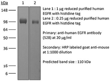
Western Blot
Bessette, D. C., et al. (2015). "Using the MCF10A/MCF10CA1a Breast Cancer Progression Cell Line Model to Investigate the Effect of Active, Mutant Forms of EGFR in Breast Cancer Development and Treatment Using Gefitinib" PLoS One 10(5): e0125232. PubMed
BACKGROUND: Basal-like and triple negative breast cancer (TNBC) share common molecular features, poor prognosis and a propensity for metastasis to the brain. Amplification of epidermal growth factor receptor (EGFR) occurs in ~50% of basal-like breast cancer, and mutations in the epidermal growth factor receptor (EGFR) have been reported in up to ~ 10% of Asian TNBC patients. In non-small cell lung cancer several different mutations in the EGFR tyrosine kinase domain confer sensitivity to receptor tyrosine kinase inhibitors, but the tumourigenic potential of EGFR mutations in breast cells and their potential for targeted therapy is unknown. MATERIALS AND METHODS: Constructs containing wild type, G719S or E746-A750 deletion mutant forms of EGFR were transfected into the MCF10A breast cells and their tumorigenic derivative, MCF10CA1a. The effects of EGFR over-expression and mutation on proliferation, migration, invasion, response to gefitinib, and tumour formation in vivo was investigated. Copy number analysis and whole exome sequencing of the MCF10A and MCF10CA1a cell lines were also performed. RESULTS: Mutant EGFR increased MCF10A and MCF10CA1a proliferation and MCF10A gefitinib sensitivity. The EGFR-E746-A750 deletion increased MCF10CA1a cell migration and invasion, and greatly increased MCF10CA1a xenograft tumour formation and growth. Compared to MCF10A cells, MCF10CA1a cells exhibited large regions of gain on chromosomes 3 and 9, deletion on chromosome 7, and mutations in many genes implicated in cancer. CONCLUSIONS: Mutant EGFR enhances the oncogenic properties of MCF10A cell line, and increases sensitivity to gefitinib. Although the addition of EGFR E746-A750 renders the MCF10CA1a cells more tumourigenic in vivo it is not accompanied by increased gefitinib sensitivity, perhaps due to additional mutations, including the PIK3CA H1047R mutation, that the MCF10CA1a cell line has acquired. Screening TNBC/basal-like breast cancer for EGFR mutations may prove useful for directing therapy but, as in non-small cell lung cancer, accompanying mutations in PIK3CA may confer gefitinib resistance.
in vivo EGFR blockade in xenografts, Western Blot
Black, P. C., et al. (2011). "Receptor heterodimerization: a new mechanism for platelet-derived growth factor induced resistance to anti-epidermal growth factor receptor therapy for bladder cancer" J Urol 185(2): 693-700. PubMed
PURPOSE: Human bladder cancer cells resistant to anti-epidermal growth factor receptor therapy often co-express platelet-derived growth factor receptor-beta. We determined whether there is functional crosstalk between epidermal growth factor receptor and platelet-derived growth factor receptor-beta, and how this regulates biological functions in bladder cancer cases. MATERIALS AND METHODS: We determined heterodimerization and co-localization of epidermal growth factor receptor and platelet-derived growth factor receptor-beta by immunoprecipitation and confocal microscopy, respectively. We tested the antiproliferative effects of specific inhibitory monoclonal antibodies to each receptor by (3)H-thymidine uptake assay. We transfected the nonplatelet-derived growth factor receptor-beta expressing bladder cancer cell line UMUC5 with the platelet-derived growth factor receptor-beta gene. These cells were analyzed in vitro by (3)H-thymidine uptake and by Matrigel invasion assay, and in vivo for tumorigenicity, metastatic potential and orthotopic growth. In a treatment study nude mice were inoculated with orthotopic tumors and treated with the inhibitory antibodies alone and in combination. RESULTS: Immunoprecipitation revealed epidermal growth factor receptor/platelet-derived growth factor receptor-beta heterodimers in all platelet-derived growth factor receptor-beta expressing cell lines. Forced expression of platelet-derived growth factor receptor-beta in epidermal growth factor receptor sensitive UMUC5 cells (50% inhibitory concentration less than 10 nM) significantly decreased responsiveness to epidermal growth factor receptor inhibition (50% inhibitory concentration greater than 100 nM) and increased invasive potential 3-fold as well as tumorigenicity. Increased invasiveness was associated with epidermal growth factor triggered platelet-derived growth factor receptor-beta transactivation, increased mitogen activated protein kinase and glycogen synthase kinase-3beta phosphorylation, and decreased E-cadherin. Inhibition of epidermal growth factor receptor and platelet-derived growth factor receptor-beta receptors blocked cell invasion, decreased cell proliferation, reduced xenograft tumor growth and increased E-cadherin expression. CONCLUSIONS: In epidermal growth factor receptor expressing bladder cancer co-expression of platelet-derived growth factor receptor-beta has implications for tumor biology. Thus, it should be further evaluated as a strategy involving dual receptor targeting.
in vitro EGFR blockade
Tokumaru, S., et al. (2005). "Induction of keratinocyte migration via transactivation of the epidermal growth factor receptor by the antimicrobial peptide LL-37" J Immunol 175(7): 4662-4668. PubMed
The closure of skin wounds is essential for resistance against microbial pathogens, and keratinocyte migration is an important step in skin wound healing. Cathelicidin hCAP18/LL-37 is an innate antimicrobial peptide that is expressed in the skin and acts to eliminate microbial pathogens. Because hCAP18/LL-37 is up-regulated at skin wound sites, we hypothesized that LL-37 induces keratinocyte migration. In this study, we found that 1 microg/ml LL-37 induced the maximum level of keratinocyte migration in the Boyden chamber assay. In addition, LL-37 phosphorylated the epidermal growth factor receptor (EGFR) after 10 min, which suggests that LL-37-induced keratinocyte migration occurs via EGFR transactivation. To test this assumption, we used inhibitors that block the sequential steps of EGFR transactivation, such as OSU8-1, CRM197, anti-EGFR no. 225 Ab, and AG1478. All of these inhibitors completely blocked LL-37-induced keratinocyte migration, which indicates that migration occurs via HB-EGF-mediated EGFR transactivation. Furthermore, CRM197, anti-EGFR no. 225, and AG1478 blocked the LL-37-induced phosphorylation of STAT3, and transfection with a dominant-negative mutant of STAT3 abolished LL-37-induced keratinocyte migration, indicating the involvement of the STAT3 pathway downstream of EGFR transactivation. Finally, we tested whether the suppressor of cytokine signaling (SOCS)/cytokine-inducible Src homology 2-containing protein (CIS) family of negative regulators of STAT3 regulates LL-37-induced keratinocyte migration. Transfection with SOCS1/Jak2 binding protein or SOCS3/CIS3 almost completely abolished LL-37-induced keratinocyte migration. In conclusion, LL-37 induces keratinocyte migration via heparin-binding-EGF-mediated transactivation of EGFR, and SOCS1/Jak 2 binding and SOCS3/CIS3 negatively regulate this migration. The results of this study suggest that LL-37 closes skin wounds by the induction of keratinocyte migration.
in vitro EGFR blockade
Lesma, E., et al. (2005). "Isolation and growth of smooth muscle-like cells derived from tuberous sclerosis complex-2 human renal angiomyolipoma: epidermal growth factor is the required growth factor" Am J Pathol 167(4): 1093-1103. PubMed
Tuberous sclerosis complex (TSC) is a tumor suppressor gene disorder characterized by mutations in the TSC1 or TSC2 genes. These mutations lead to the development of benign tumors involving smooth muscle cells, causing life-threatening lymphangioleiomyomatosis. We isolated and characterized two types of cells bearing a mutation in TSC2 exon 18 from a renal angiomyolipoma of a TSC patient: one population of alpha-actin-positive smooth muscle-like cells with loss of heterozygosity for the TSC2 gene (A(+) cells) and another of nonloss of heterozygosity keratin 8/18-positive epithelial-like cells (R(+) cells). Unlike control aortic vascular smooth muscle cells, A(+) cells required epidermal growth factor (EGF) to grow and substituting EGF with insulin-like growth factor (IGF)-1 failed to increase the cell number; however, omission of EGF did not cause cell loss. The A(+) cells constantly released IGF-1 into the culture medium and constitutively showed a high degree of S6K phosphorylation even when grown in serum-free medium. Exposure to antibodies against EGF and IGF-1 receptors caused a rapid loss of A(+) cells: 50% by 5 days and 100% by 12 days. Signal transduction mediated by EGF and IGF-I receptors is therefore involved in A(+) cell survival. These results may offer a novel therapeutic perspective for the treatment of TSC complications and lymphangioleiomyomatosis.
in vitro EGFR blockade
Sherwood, E. R., et al. (1998). "Epidermal growth factor receptor activation in androgen-independent but not androgen-stimulated growth of human prostatic carcinoma cells" Br J Cancer 77(6): 855-861. PubMed
These studies were undertaken to assess the relative expression and autocrine activation of the epidermal growth factor receptor (EGFR) in normal and transformed prostatic epithelial cells and to determine whether EGFR activation plays a functional role in androgen-stimulated growth of prostate cancer cells in vitro. EGFR expression was determined by Western blot analysis and ELISA immunoassays. Immunoprecipitation of radiophosphorylated EGFR and evaluation of tyrosine phosphorylation was used to assess EGFR activation. The human androgen-independent prostate cancer cell lines PC3 and DU145 exhibited higher levels of EGFR expression and autocrine phosphorylation than normal human prostatic epithelial cells or the human androgen-responsive prostate cancer cell line LNCaP. PC3 and DU145 cells also showed higher levels of autonomous growth under serum-free defined conditions. Normal prostatic epithelial cells expressed EGFR but did not exhibit detectable levels of EGFR phosphorylation when cultured in the absence of exogenous EGF. Addition of EGF stimulated EGFR phosphorylation and induced proliferation of normal cells. LNCaP cells exhibited autocrine phosphorylation of EGFR but did not undergo significant proliferation when cultured in the absence of exogenous growth factors. A biphasic growth curve was observed when LNCaP cells were cultured with dihydrotestosterone (DHT). Maximum proliferation occurred at 1 nM DHT with regression of the growth response at DHT concentrations greater than 1 nM. However, neither EGFR expression nor phosphorylation was altered in LNCaP cells after androgen stimulation. In addition, DHT-stimulated growth of LNCaP cells was not inhibited by anti-EGFR. These studies show that autocrine activation of EGFR is a common feature of prostatic carcinoma cells in contrast to normal epithelial cells. However, EGFR activation does not appear to play a functional role in androgen-stimulated growth of LNCaP cells in vitro.
Functional Assays
Kawamoto, T., et al. (1983). "Growth stimulation of A431 cells by epidermal growth factor: identification of high-affinity receptors for epidermal growth factor by an anti-receptor monoclonal antibody" Proc Natl Acad Sci U S A 80(5): 1337-1341. PubMed
Epidermal growth factor (EGF) at 3 nM maximally inhibits the proliferation of A431 epidermoid carcinoma cells. We show that at lower concentrations, in the range of 3-100 pM, EGF has a mitogenic effect on A431 cells. In the presence of 100 nM anti-EGF-receptor monoclonal IgG (designated 528), which inhibits A431 cell proliferation and blocks greater than 95% of EGF binding, EGF becomes mitogenic for A431 cells at concentrations up to 3 nM. These results suggest that a minor population of high-affinity EGF receptors may be involved in stimulation of A431 cell proliferation. Saturation binding assays with 125I-labeled EGF indicate that approximately equal to 0.1-0.2% of receptors for EGF are high-affinity receptors that bind EGF with an estimated Kd of 7 X 10(-11) M. This affinity is nearly 2 orders of magnitude higher than that of the remaining EGF receptors. Although A431 cell proliferation is maximally inhibited by nonsaturating amounts of EGF (3 nM), maximal inhibition by 528 IgG (approximately equal to 70% of maximal inhibition by EGF) requires saturating concentrations of antibody (approximately equal to 15 nM). Unlike EGF, rapid down-regulation is not observed with 528 IgG. These results indicate different mechanisms of growth inhibition of A431 cells by EGF and 528 IgG.