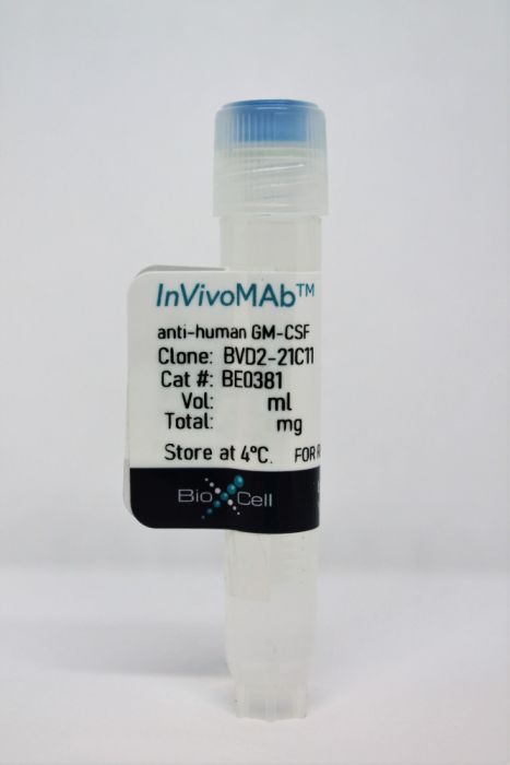
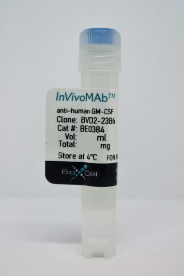
InVivoMAb anti-human GM-CSF
Product Details
The BVD2-21C11 monoclonal antibody reacts with human granulocyte-macrophage colony-stimulating factor (GM-CSF), also known as colony stimulating factor 2 (CSF2). GM-CSF is a 14 kDa monomeric hematopoietic factor secreted by macrophages, T cells, mast cells, NK cells, endothelial cells, and fibroblasts. GM-CSF stimulates stem cells to differentiate into granulocytes (neutrophils, eosinophils, and basophils) and monocytes. The BVD2-21C11 antibody has been reported to cross-react with GM-CSF from the rhesus monkey. BVD2-21C11 has been reported as a neutralizing antibody.Specifications
| Isotype | Rat IgG2a, κ |
|---|---|
| Recommended Isotype Control(s) | InVivoMAb rat IgG2a isotype control, anti-trinitrophenol |
| Recommended Dilution Buffer | InVivoPure pH 7.0 Dilution Buffer |
| Conjugation | This product is unconjugated. Conjugation is available via our Antibody Conjugation Services. |
| Immunogen | Recombinant human GM-CSF |
| Reported Applications |
Immunofluorescence ELISA Flow cytometry |
| Formulation |
PBS, pH 7.0 Contains no stabilizers or preservatives |
| Endotoxin |
<2EU/mg (<0.002EU/μg) Determined by LAL gel clotting assay |
| Purity |
>95% Determined by SDS-PAGE |
| Sterility | 0.2 µm filtration |
| Purification | Protein G |
| RRID | AB_2927518 |
| Molecular Weight | 150 kDa |
| Storage | The antibody solution should be stored at the stock concentration at 4°C. Do not freeze. |
Recommended Products
Flow Cytometry
Pearson, C., et al. (2016). "ILC3 GM-CSF production and mobilisation orchestrate acute intestinal inflammation" Elife 5: e10066. PubMed
Innate lymphoid cells (ILCs) contribute to host defence and tissue repair but can induce immunopathology. Recent work has revealed tissue-specific roles for ILCs; however, the question of how a small population has large effects on immune homeostasis remains unclear. We identify two mechanisms that ILC3s utilise to exert their effects within intestinal tissue. ILC-driven colitis depends on production of granulocyte macrophage-colony stimulating factor (GM-CSF), which recruits and maintains intestinal inflammatory monocytes. ILCs present in the intestine also enter and exit cryptopatches in a highly dynamic process. During colitis, ILC3s mobilize from cryptopatches, a process that can be inhibited by blocking GM-CSF, and mobilization precedes inflammatory foci elsewhere in the tissue. Together these data identify the IL-23R/GM-CSF axis within ILC3 as a key control point in the accumulation of innate effector cells in the intestine and in the spatio-temporal dynamics of ILCs in the intestinal inflammatory response.
Flow Cytometry
Kaufmann, U., et al. (2016). "Selective ORAI1 Inhibition Ameliorates Autoimmune Central Nervous System Inflammation by Suppressing Effector but Not Regulatory T Cell Function" J Immunol 196(2): 573-585. PubMed
The function of CD4(+) T cells is dependent on Ca(2+) influx through Ca(2+) release-activated Ca(2+) (CRAC) channels formed by ORAI proteins. To investigate the role of ORAI1 in proinflammatory Th1 and Th17 cells and autoimmune diseases, we genetically and pharmacologically modulated ORAI1 function. Immunization of mice lacking Orai1 in T cells with MOG peptide resulted in attenuated severity of experimental autoimmune encephalomyelitis (EAE). The numbers of T cells and innate immune cells in the CNS of ORAI1-deficient animals were strongly reduced along with almost completely abolished production of IL-17A, IFN-gamma, and GM-CSF despite only partially reduced Ca(2+) influx. In Th1 and Th17 cells differentiated in vitro, ORAI1 was required for cytokine production but not the expression of Th1- and Th17-specific transcription factors T-bet and RORgammat. The differentiation and function of induced regulatory T cells, by contrast, was independent of ORAI1. Importantly, induced genetic deletion of Orai1 in adoptively transferred, MOG-specific T cells was able to halt EAE progression after disease onset. Likewise, treatment of wild-type mice with a selective CRAC channel inhibitor after EAE onset ameliorated disease. Genetic deletion of Orai1 and pharmacological ORAI1 inhibition reduced the leukocyte numbers in the CNS and attenuated Th1/Th17 cell-mediated cytokine production. In human CD4(+) T cells, CRAC channel inhibition reduced the expression of IL-17A, IFN-gamma, and other cytokines in a dose-dependent manner. Taken together, these findings support the conclusion that Th1 and Th17 cell function is particularly dependent on CRAC channels, which could be exploited as a therapeutic approach to T cell-mediated autoimmune diseases.
Immunofluorescence, Flow Cytometry
Rasouli, J., et al. (2015). "Expression of GM-CSF in T Cells Is Increased in Multiple Sclerosis and Suppressed by IFN-beta Therapy" J Immunol 194(11): 5085-5093. PubMed
Multiple sclerosis (MS) is an autoimmune disease of the CNS. Studies in animal models of MS have shown that GM-CSF produced by T cells is necessary for the development of autoimmune CNS inflammation. This suggests that GM-CSF may have a pathogenic role in MS as well, and a clinical trial testing its blockade is ongoing. However, there have been few reports on GM-CSF production by T cells in MS. The objective of this study was to characterize GM-CSF production by T cells of MS patients and to determine the effect of IFN-beta therapy on its production. GM-CSF production by peripheral blood (PB) T cells and the effects of IFN-beta were characterized in samples of untreated and IFN-beta-treated MS patients versus healthy subjects. GM-CSF production by T cells in MS brain lesions was analyzed by immunofluorescence. Untreated MS patients had significantly greater numbers of GM-CSF(+)CD4(+) and CD8(+) T cells in PB compared with healthy controls and IFN-beta-treated MS patients. IFN-beta significantly suppressed GM-CSF production by T cells in vitro. A number of CD4(+) and CD8(+) T cells in MS brain lesions expressed GM-CSF. Elevated GM-CSF production by PB T cells in MS is indicative of aberrant hyperactivation of the immune system. Given its essential role in animal models, abundant GM-CSF production at the sites of CNS inflammation suggests that GM-CSF contributes to MS pathogenesis. Our findings also reveal a potential mechanism of IFN-beta therapy, namely suppression of GM-CSF production.
Flow Cytometry
Li, R., et al. (2015). "Proinflammatory GM-CSF-producing B cells in multiple sclerosis and B cell depletion therapy" Sci Transl Med 7(310): 310ra166. PubMed
B cells are not limited to producing protective antibodies; they also perform additional functions relevant to both health and disease. However, the relative contribution of functionally distinct B cell subsets in human disease, the signals that regulate the balance between such subsets, and which of these subsets underlie the benefits of B cell depletion therapy (BCDT) are only partially elucidated. We describe a proinflammatory, granulocyte macrophage-colony stimulating factor (GM-CSF)-expressing human memory B cell subset that is increased in frequency and more readily induced in multiple sclerosis (MS) patients compared to healthy controls. In vitro, GM-CSF-expressing B cells efficiently activated myeloid cells in a GM-CSF-dependent manner, and in vivo, BCDT resulted in a GM-CSF-dependent decrease in proinflammatory myeloid responses of MS patients. A signal transducer and activator of transcription 5 (STAT5)- and STAT6-dependent mechanism was required for B cell GM-CSF production and reciprocally regulated the generation of regulatory IL-10-expressing B cells. STAT5/6 signaling was enhanced in B cells of untreated MS patients compared with healthy controls, and B cells reemerging in patients after BCDT normalized their STAT5/6 signaling as well as their GM-CSF/IL-10 cytokine secretion ratios. The diminished proinflammatory myeloid cell responses observed after BCDT persisted even as new B cells reconstituted. These data implicate a proinflammatory B cell/myeloid cell axis in disease and underscore the rationale for selective targeting of distinct B cell populations in MS and other human autoimmune diseases.
ELISA
Kitamura, T., et al. (1999). "Idiopathic pulmonary alveolar proteinosis as an autoimmune disease with neutralizing antibody against granulocyte/macrophage colony-stimulating factor" J Exp Med 190(6): 875-880. PubMed
Idiopathic pulmonary alveolar proteinosis (I-PAP) is a rare disease of unknown etiology in which the alveoli fill with lipoproteinaceous material. We report here that I-PAP is an autoimmune disease with neutralizing antibody of immunoglobulin G isotype against granulocyte/macrophage colony-stimulating factor (GM-CSF). The antibody was found to be present in all specimens of bronchoalveolar lavage fluid obtained from 11 I-PAP patients but not in samples from 2 secondary PAP patients, 53 normal subjects, and 14 patients with other lung diseases. It specifically bound GM-CSF and neutralized bioactivity of the cytokine in vitro. The antibody was also found in sera from all I-PAP patients examined but not in sera from a secondary PAP patient or normal subjects, indicating that it exists systemically in I-PAP patients. As lack of GM-CSF signaling causes PAP in congenital cases and PAP-like disease in murine models, our findings strongly suggest that neutralization of GM-CSF bioactivity by the antibody causes dysfunction of alveolar macrophages, which results in reduced surfactant clearance.


