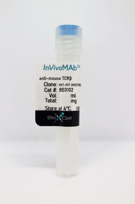
InVivoMAb anti-mouse TCRβ
Product Details
The H57-597 monoclonal antibody reacts with the beta chain of the mouse T cell receptor (TCR). TCRβ belongs to the immunoglobulin superfamily and is expressed by thymocytes and T lymphocytes. TCRβ combines with TCRα to form TCR α/β. TCR α/β plays a central role in antigen recognition, signal transduction, and T cell activation. The H57-597 antibody has been shown to deplete TCR α/β bearing T cells following in vivo administration.Specifications
| Isotype | Armenian hamster IgG |
|---|---|
| Recommended Isotype Control(s) | InVivoMAb polyclonal Armenian hamster IgG |
| Recommended Dilution Buffer | InVivoPure pH 7.0 Dilution Buffer |
| Conjugation | This product is unconjugated. Conjugation is available via our Antibody Conjugation Services. |
| Immunogen | Affinity purified TCR from mouse DO-11.10 cells |
| Reported Applications | in vivo T cell depletion |
| Formulation |
PBS, pH 7.0 Contains no stabilizers or preservatives |
| Endotoxin |
<2EU/mg (<0.002EU/μg) Determined by LAL gel clotting assay |
| Purity |
>95% Determined by SDS-PAGE |
| Sterility | 0.2 µm filtration |
| Production | Purified from cell culture supernatant in an animal-free facility |
| Purification | Protein G |
| RRID | AB_10950158 |
| Molecular Weight | 150 kDa |
| Storage | The antibody solution should be stored at the stock concentration at 4°C. Do not freeze. |
Recommended Products
-
Recommended Isotype Control(s)
InVivoMAb polyclonal Armenian hamster IgG
-

Recommended Dilution Buffer
InVivoPure pH 7.0 Dilution Buffer
in vitro T cell stimulation/activation
Kimura, S., et al. (2019). "Sox8 is essential for M cell maturation to accelerate IgA response at the early stage after weaning in mice" J Exp Med 216(4): 831-846. PubMed
Microfold (M) cells residing in the follicle-associated epithelium (FAE) of the gut-associated lymphoid tissue are specialized for antigen uptake to initiate mucosal immune responses. The molecular machinery and biological significance of M cell differentiation, however, remain to be fully elucidated. Here, we demonstrate that Sox8, a member of the SRY-related HMG box transcription factor family, is specifically expressed by M cells in the intestinal epithelium. The expression of Sox8 requires activation of RANKL-RelB signaling. Chromatin immunoprecipitation and luciferase assays revealed that Sox8 directly binds the promoter region of Gp2 to increase Gp2 expression, which is the hallmark of functionally mature M cells. Furthermore, genetic deletion of Sox8 causes a marked decrease in the number of mature M cells, resulting in reduced antigen uptake in Peyer’s patches. Consequently, juvenile Sox8-deficient mice showed attenuated germinal center reactions and antigen-specific IgA responses. These findings indicate that Sox8 plays an essential role in the development of M cells to establish mucosal immune responses.
in vivo T cell depletion
Emgård, J., et al. (2018). "Oxysterol Sensing through the Receptor GPR183 Promotes the Lymphoid-Tissue-Inducing Function of Innate Lymphoid Cells and Colonic Inflammation" Immunity 48(1): 120-132.e128. PubMed
Group 3 innate lymphoid cells (ILC3s) sense environmental signals and are critical for tissue integrity in the intestine. Yet, which signals are sensed and what receptors control ILC3 function remain poorly understood. Here, we show that ILC3s with a lymphoid-tissue-inducer (LTi) phenotype expressed G-protein-coupled receptor 183 (GPR183) and migrated to its oxysterol ligand 7α,25-hydroxycholesterol (7α,25-OHC). In mice lacking Gpr183 or 7α,25-OHC, ILC3s failed to localize to cryptopatches (CPs) and isolated lymphoid follicles (ILFs). Gpr183 deficiency in ILC3s caused a defect in CP and ILF formation in the colon, but not in the small intestine. Localized oxysterol production by fibroblastic stromal cells provided an essential signal for colonic lymphoid tissue development, and inflammation-induced increased oxysterol production caused colitis through GPR183-mediated cell recruitment. Our findings show that GPR183 promotes lymphoid organ development and indicate that oxysterol-GPR183-dependent positioning within tissues controls ILC3 activity and intestinal homeostasis.
in vivo T cell depletion
Schroder, P. M., et al. (2013). "Transient combination therapy targeting the immune synapse abrogates T cell responses and prolongs allograft survival in mice" PLoS One 8(7): e69397. PubMed
T cells play a major role in allograft rejection, which occurs after T cell activation by the engagement of several functional molecules to form an immune synapse with alloantigen presenting cells. In this study, the immune synapse was targeted using mAbs directed to the TCR beta-chain (TCRbeta) and lymphocyte function-associated antigen-1 (LFA1) to induce long-term allograft survival. Evaluation of antigen-specific T cell responses was performed by adoptively transferring CFSE labeled transgenic OT-II cells into wild-type mice and providing OVA peptide by intravenous injection. Graft survival studies were performed in mice by transplanting BALB/c ear skins onto the flanks of C57BL/6 recipients. The anti-TCRbeta plus anti-LFA1 mAb combination (but not either mAb alone) abrogated antigen-specific T cell responses invitro and invivo. Transient combination therapy with these agents resulted in significantly prolonged skin allograft survival in mice (51+/-10 days; p<0.01) when compared to treatment with either anti-TCRbeta mAb (24+/-5 days) or anti-LFA1 mAb (19+/-3 days) alone or no treatment (10+/-1 days). When lymphoid tissues from these mice were analyzed at different times post-transplant, only those receiving the combination of anti-TCRbeta and anti-LFA1 mAbs demonstrated long-lasting reductions in total T cell numbers, cellular and humoral anti-donor responses, and expression of CD3 on the surface of T cells. These results demonstrate that transient anti-TCRbeta and anti-LFA1 mAb combination therapy abrogates antigen-reactive T cell responses with long-lasting effects that significantly prolong allograft survival.
in vivo T cell depletion
Gillard, G. O., et al. (2011). "Thy1+ NK cells from vaccinia virus-primed mice confer protection against vaccinia virus challenge in the absence of adaptive lymphocytes" PLoS Pathog 7(8): e1002141. PubMed
While immunological memory has long been considered the province of T- and B-lymphocytes, it has recently been reported that innate cell populations are capable of mediating memory responses. We now show that an innate memory immune response is generated in mice following infection with vaccinia virus, a poxvirus for which no cognate germline-encoded receptor has been identified. This immune response results in viral clearance in the absence of classical adaptive T and B lymphocyte populations, and is mediated by a Thy1(+) subset of natural killer (NK) cells. We demonstrate that immune protection against infection from a lethal dose of virus can be adoptively transferred with memory hepatic Thy1(+) NK cells that were primed with live virus. Our results also indicate that, like classical immunological memory, stronger innate memory responses form in response to priming with live virus than a highly attenuated vector. These results demonstrate that a defined innate memory cell population alone can provide host protection against a lethal systemic infection through viral clearance.